Quantcast
Channel: cholesterol – Jeffrey Dach MD
Viewing all 55 articles
Browse latest View live

A Neurosurgeon with a Painful Arm, Myositis from Statin and PPI Drugs by Jeffrey Dach MD


Statin Nation, The Movie

$
0
0

Statin Nation, the full length movie will inform you about cholesterol and cholesterol lowering drugs. Statin cholesterol lowering drugs work for a subset of the population at reducing risk for heart disease.  However, for most healthy people, statin drugs are … Continue reading

The post Statin Nation, The Movie appeared first on Jeffrey Dach MD .

Familial Hypercholesterolemia and Statin Drugs by Jeffrey Dach MD

Atherosclerosis and Heart Disease How Does it Happen

Reversing Heart Disease without Drugs

$
0
0

Reversing Heart Disease without Drugs.  Heart Disease is today’s number one killer in America and is the reason why 16 million Americans are taking a Statin anti-cholesterol drug.  This article explains what causes heart disease… Read More

The post Reversing Heart Disease without Drugs appeared first on Jeffrey Dach MD .

Does High Cholesterol Cause Heart Disease ?

Healthy Men Should Not Take Statin Drugs by Jeffrey Dach MD

The Art of the Curb Side Cholesterol Consult by Jeffrey Dach MD


Lipitor and The Dracula of Modern Technology

Defending the Cholesterol Hypothesis in the Elderly

$
0
0

MedPage_Today Defending the Cholesterol Hypothesis in the Elderly

by Jeffrey Dach MD

Med Page Today ran an article by cardiologist Larry Husten, defending the cholesterol theory of heart disease entitled “Cholesterol Skeptics Launch Another Attack”. (1)(10)  Left Image Medpage Today Logo courtesy of Medpage Today.

Uffe Ravnskov Cholesterol Heart Disease StatinDr Husten is responding to an article published in British Medical Journal by Dr Uffe Ravnskov (left image) in which a number of previously published studies contradict the cholesterol theory by showing an “Inverse association between low-density-lipoprotein (LDL) cholesterol and mortality in the elderly”.(2) 

In other words,  higher serum cholesterol in the elderly is associated with decreased mortality.   This directly contradicts the cholesterol theory which states that higher cholesterol should increase mortality from heart disease.  In the elderly, it doesn’t work this way.  Old people tend to live longer if they have a higher serum cholesterol.  Low serum cholesterol in the elderly is a marker for increased mortality.

Cholesterol Theory has Been Falsified

Current accepted dogma proposes cholesterol as the cause of  atherosclerotic plaque, and therefore, reduction of serum cholesterol with a statin drug (left image)  should prevent coronary artery disease. Quite to the contrary, the cholesterol theory has been falsified. According to Dr. William R Ware, there is no correlation between serum cholesterol and the amount of atherosclerotic plaque when reviewing either autopsy studies or coronary calcium score studies.(5)

The lack of relationship between serum cholesterol and calcium score was reported by Dr. Hecht who says:

”There were no significant differences in the calcium scores throughout the entire range of all lipid parameters; calcium percentiles were virtually identical within lipid value subgroups.”(6)

More than a dozen studies show low cholesterol in the elderly is a marker for increased mortality, not improved survival.  In women, cholesterol lowering drugs should not be used as they do not improve survival and do not improve health.

cholesterol Drug AdAlso, in selected medical conditions such as congestive heart failure, haemodialysis, chronic obstructive pulmonary disease (COPD), as in the elderly, higher cholesterol is associated with improved survival, and lower cholesterol with increased mortality.(9) Left Image advertising for the cholesterol theory of heart disease, creating irrational fear .  This has been falsified.

Conclusion:  Med Page Today cardiologist Larry Husten’s misplaced enthusiasm defending the cholesterol theory of heart disease is a sad commentary on the state of cardiology today.  By now, the published data is overwhelming that the cholesterol theory of heart disease has been falsified.

Jeffrey Dach MD

Articles with related Interest:

Low Level Endotoxemia Coronary Artery Disease

Getting Off Statin Drug Stories

The Art of the Curbside Cholesterol Consult

Cholesterol Lowering Drugs in the Elderly, Bad Idea

Cholesterol Lowering Drugs For Women, Just Say No.

Links and References:

1) CardioBrief: Cholesterol Skeptics Launch Another Attack
But most experts say new study is highly flawed by Larry Husten Cardiologist

Michael Blaha, MD, MPH, also of Johns Hopkins, said that he agrees with the authors that LDL by itself is a poor marker of risk in the elderly
According to Blaha, the fact that the role of LDL diminishes with age “has been known for a long time — and in fact is baked into equations like the Framingham Risk Score and the Pooled Cohort Equations.” One reason is that “cumulative exposure to high cholesterol is likely what matters most, thus very high LDL matters more in younger patients, while late-life elevation matters very little.”
In addition, Blaha said, “one must also account for survival bias in these studies,” as the older patients with high LDL are a selected group.

2) BMJ Open 2016;6:e010401 doi:10.1136/bmjopen-2015-010401
Cardiovascular medicine
Lack of an association or an inverse association between low-density-lipoprotein cholesterol and mortality in the elderly: a systematic review
Uffe Ravnskov1, David M Diamond2, Rokura Hama3, Tomohito Hamazaki4, Björn Hammarskjöld5, Niamh Hynes6, Malcolm Kendrick7, Peter H Langsjoen8, Aseem Malhotra9, Luca Mascitelli10, Kilmer S McCully11, Yoichi Ogushi12, Harumi Okuyama13, Paul J Rosch14, Tore Schersten15, Sherif Sultan6, Ralf Sundberg16

Abstract
Objective It is well known that total cholesterol becomes less of a risk factor or not at all for all-cause and cardiovascular (CV) mortality with increasing age, but as little is known as to whether low-density lipoprotein cholesterol (LDL-C), one component of total cholesterol, is associated with mortality in the elderly, we decided to investigate this issue.
Setting, participants and outcome measures We sought PubMed for cohort studies, where LDL-C had been investigated as a risk factor for all-cause and/or CV mortality in individuals ≥60 years from the general population.
Results We identified 19 cohort studies including 30 cohorts with a total of 68 094 elderly people, where all-cause mortality was recorded in 28 cohorts and CV mortality in 9 cohorts. Inverse association between all-cause mortality and LDL-C was seen in 16 cohorts (in 14 with statistical significance) representing 92% of the number of participants, where this association was recorded. In the rest, no association was found. In two cohorts, CV mortality was highest in the lowest LDL-C quartile and with statistical significance; in seven cohorts, no association was found.
Conclusions High LDL-C is inversely associated with mortality in most people over 60 years. This finding is inconsistent with the cholesterol hypothesis (ie, that cholesterol, particularly LDL-C, is inherently atherogenic). Since elderly people with high LDL-C live as long or longer than those with low LDL-C, our analysis provides reason to question the validity of the cholesterol hypothesis. Moreover, our study provides the rationale for a re-evaluation of guidelines recommending pharmacological reduction of LDL-C in the elderly as a component of cardiovascular disease prevention strategies.

3) Uffe Ravnskov The Cholesterol Myths by UffeRavnskov, MD, PhD

4) THINCS For decades, enormous human and financial resources have been wasted on the cholesterol campaign, more promising research areas have been neglected, producers and manufacturers of animal food all over the world have suffered economically, and millions of healthy people have been frightened and badgered into eating a tedious and flavorless diet or into taking potentially dangerous drugs for the rest of their lives. As the scientific evidence in support of the cholesterol campaign is non-existent, we consider it important to stop it as soon as possible.
The International Network of Cholesterol Skeptics (THINCS) is a steadily growing group of scientists, physicians, other academicians and science writers from various countries. Members of this group represent different views about the causation of atherosclerosis and cardiovascular disease, some of them are in conflict with others, but this is a normal part of science. What we all oppose is that animal fat and high cholesterol play a role. The aim with this website is to inform our colleagues and the public that this idea is not supported by scientific evidence; in fact, for many years a huge number of scientific studies have directly contradicted it.

5) Ware WR. The mainstream hypothesis that LDL cholesterol drives atherosclerosis may have been falsified by non-invasive imaging of coronary artery plaque burden and progression. Med Hypotheses. 2009;73(4):596-600.  cholesterol atherosclerosis falsified coronary artery plaque Ware Medical Hypotheses 2009

6) Hecht HS, Superko HR, Smith LK, McColgan BP. Relation of coronary artery calcium identified by electron beam tomography to serum lipoprotein levels and implications for treatment. Am J Cardiol. 2001;87(4):406-12.

7) Ravnskov U, McCully KS. Biofilms, lipoprotein aggregates, homocysteine, and arterial plaque rupture. MBio. 2014;5(5):e01717-14.Biofilms Arterial Plaque Rupture Ravnskov Uffe Kilmer McCully 2014

8) Ravnskov U, McCully KS. Infections may be causal in the pathogenesis of atherosclerosis. Am J Med Sci. 2012;344(5):391-4.  Infections May be Causal in the Pathogenesis of Atherosclerosis Ravnskov McCully 2012

9) Sandek A., Utchill S, Rauchhaus M. The endotoxin-lipoprotein hypothesis-an update. Arch Med Sci. 2007;3(4A):S81.   The endotoxin lipoprotein hypothesis Anja Sandek 2007

10)  CEBM response: “Lack of an association or an inverse association between low-density-lipoprotein cholesterol and mortality in the elderly: a systematic review” – a post publication peer review
Here we provide a post publication critical appraisal of the methodology and evidence used to support the findings from the Ravnskov et al paper.

Therefore their search strategy and reporting thereof presents a high risk of bias for missing important and relevant studies.

link to this article: http://wp.me/p3gFbV-3JT

Jeffrey Dach MD
7450 Griffin Road Suite 190
Davie, Fl 33314
954-792-4663
www.jeffreydachmd.com
http://www.drdach.com
http://www.naturalmedicine101.com
http://www.truemedmd.com

Disclaimer click here: http://www.drdach.com/wst_page20.html

The reader is advised to discuss the comments on these pages with his/her personal physicians and to only act upon the advice of his/her personal physician. Also note that concerning an answer which appears as an electronically posted question, I am NOT creating a physician — patient relationship. Although identities will remain confidential as much as possible, as I can not control the media, I can not take responsibility for any breaches of confidentiality that may occur.

Copyright (c) 2016 Jeffrey Dach MD All Rights Reserved. This article may be reproduced on the internet without permission, provided there is a link to this page and proper credit is given.

FAIR USE NOTICE: This site contains copyrighted material the use of which has not always been specifically authorized by the copyright owner. We are making such material available in our efforts to advance understanding of issues of significance. We believe this constitutes a ‘fair use’ of any such copyrighted material as provided for in section 107 of the US Copyright Law. In accordance with Title 17 U.S.C. Section 107, the material on this site is distributed without profit to those who have expressed a prior interest in receiving the included information for research and educational purposes.

Serving Areas of: Hollywood, Aventura, Miami, Fort Lauderdale, Pembroke Pines, Miramar, Davie, Coral Springs, Cooper City, Sunshine Ranches, Hallandale, Surfside, Miami Beach, Sunny Isles, Normandy Isles, Coral Gables, Hialeah, Golden Beach ,Kendall,sunrise, coral springs, parkland,pompano, boca raton, palm beach, weston, dania beach, tamarac, oakland park, boynton beach, delray,lake worth,wellington,plantation.

The post Defending the Cholesterol Hypothesis in the Elderly appeared first on Jeffrey Dach MD.

Statins Reduce Peri-Operative Mortality Surely You Must Be Joking

$
0
0

Statins Reduce Peri-Operative Mortality, Surely You Must Be Joking

Another Medical Hoax

I was intrigued by Dr London’s report in December 2016 JAMA of decreased peri-operative mortality in statin users.(1)  This result does not seem immediately intuitive.  Statin drugs inhibit an enzyme in the liver called HMG-CoA reductase involved in cholesterol production, thereby reducing cholesterol levels.  Left Image: Surgeons by Reginald Brill 1934 courtesy of Wikimedia Commons.

Higher Cholesterol is Protective of Post Operative Sepsis

Higher cholesterol in the surgical patient is protective of endotoxemia, a dreaded complication of non-cardiac surgery which carries a high mortality rate. According to Dr Wilson writing in Critical Care 2003, ” lipoproteins can bind and neutralize lipopolysaccharide, hypocholesterolemia can negatively impact outcome.” (2)

Severly ILL Patients Have Low Cholesterol

Another observation made by Dr Wilson is that severely ill patients tend to have low cholesterol. If so, then these severely ill patients would not be good candidates for statin drugs in the days prior to their surgical procedure as their cholesterol levels are already low. No need to give a drug to make it lower. On the other hand non-critically ill patients tend to have higher cholesterol values which does make them good candidates for a statin drug.

The Cholesterol Endotoxin Hypothesis

This explains the spurious findings of the JAMA article. The patients not taking statins were sicker with lower cholesterol values, a marker for increased mortality from sepsis.

Conclusion:

Low cholesterol is an excellent marker for increased mortality from sepsis. (3-7)  Therefore driving down serum cholesterol with a statin drug is not a way to reduce surgical complications, most of which are related to post op sepsis.  This Study in JAMA by Dr London is therefore a medical hoax, a result of patient selection bias, not due to any imaginary benefits of statin drugs, which are non-existent.

The damage of this JAMA article could produce is frightening, since some surgeons may actually believe it and give their patients statin drugs.  This would be a catastrophe of monumental proportions.

This article is part one, for part two click here,

Jeffrey Dach MD

Articles with Related Interest:

Statin Drugs and Botanicals Anti-Inflammatory Effects

Low Level Endotoxemia and Cholesterol

Jeffrey Dach MD

Links and References:

1) Association of Perioperative Statin Use With Mortality and Morbidity After Major Noncardiac Surgery. Martin J London, JAMA Dec 19 2016.

Question Is exposure to a statin in the early perioperative period associated with reduced postoperative complications after noncardiac surgery?

Findings This observational cohort analysis of veterans linked risk and outcome data from the Veterans Affairs Surgical Quality Improvement Program database to statin prescriptions in 180 478 patients and evaluated the associations of early statin exposure on 30-day mortality. After adjustment for risk, other medications used, and potential selection biases, 30-day mortality was significantly reduced in the statin-exposed group.

Meaning Perioperative statin use may be beneficial in reducing 30-day mortality, although the effects of selection biases cannot be excluded.
Abstract

Importance The efficacy of statins in reducing perioperative cardiovascular and other organ system complications in patients undergoing noncardiac surgery remains controversial. Owing to a paucity of randomized clinical trials, analyses of large databases may facilitate informed hypothesis generation and more efficient trial design.

Objective To evaluate associations of early perioperative statin use with outcomes in a national cohort of veterans undergoing noncardiac surgery.

Design, Setting, and Participants This retrospective, observational cohort analysis included 180 478 veterans undergoing elective or emergent noncardiac surgery (including vascular, general, neurosurgery, orthopedic, thoracic, urologic, and otolaryngologic) who were admitted within 7 days of surgery and sampled by the Veterans Affairs Surgical Quality Improvement Program (VASQIP). Patients were admitted to Department of Veterans Affairs hospitals and underwent 30-day postoperative follow-up. Data were collected from October 1, 2005, to September 30, 2010, and analyzed from November 28, 2013, to October 31, 2016.

Exposure Statin use on the day of or the day after surgery.

Main Outcomes and Measures All-cause 30-day mortality (primary outcome) and standardized 30-day cardiovascular and noncardiovascular outcomes captured by VASQIP. Use of statins and other perioperative cardiovascular medications was ascertained from the Veterans Affairs Pharmacy Benefits Management research database.

Results A total of 180 478 eligible patients (95.6% men and 4.4% women; mean [SD] age, 63.8 [11.6] years) underwent analysis, and 96 486 were included in the propensity score–matched cohort (96.3% men; 3.7% women; mean [SD] age, 65.9 [10.6] years). At the time of hospital admission, 37.8% of patients had an active outpatient prescription for a statin, of whom 80.8% were prescribed simvastatin and 59.5% used moderate-intensity dosing. Exposure to a statin on the day of or the day after surgery based on an inpatient prescription was noted in 31.5% of the cohort. Among 48 243 propensity score–matched pairs of early perioperative statin-exposed and nonexposed patients, 30-day all-cause mortality was significantly reduced in exposed patients (relative risk, 0.82; 95% CI, 0.75-0.89; P < .001; number needed to treat, 244; 95% CI, 170-432). Of the secondary outcomes, a significant association with reduced risk of any complication was noted (relative risk, 0.82; 95% CI, 0.79-0.86; P < .001; number needed to treat, 67; 95% CI, 55-87); all were significant except for the central nervous system and thrombosis categories, with the greatest risk reduction (relative risk, 0.73; 95% CI, 0.64-0.83) for cardiac complications.

Conclusions and Relevance: Early perioperative exposure to a statin was associated with a significant reduction in all-cause perioperative mortality and several cardiovascular and noncardiovascular complications. However, the potential for selection biases in these results must be considered.

2) Wilson, Robert F., Jeffrey F. Barletta, and James G. Tyburski. “Hypocholesterolemia in sepsis and critically ill or injured patients.” Critical Care 7.6 (2003): 1.

3) Leardi, S., et al. “[Blood levels of cholesterol and postoperative septic complications].” Annali italiani di chirurgia 71.2 (1999): 233-237.
Hypocholesterolemia seems to represent a significant predictive factor of morbidity and mortality in critically ill patients. The authors, on the basis of recent literature data, aim to clarify the possible correlation between preoperative hypocholesterolemia and the risk of septic postoperative complications .205 patients undergoing to surgery for gastrointestinal diseases were the object of the study. Patients undergoing “minor” abdominal surgery or video-laparoscopic surgery and classified ASA III-IV were excluded. In all the patients, we considered retrospectively risk factors for postoperative septic complications as follows: preoperative blood concentration of cholesterol, malnutrition, obesity, diabetes, neoplasm, preoperative sepsis, type and duration of operations, antibiotics and regimen of use. Type and incidence of postoperative local or systemic septic complications were recorded. The patients have been stratified according to blood concentration of cholesterol and to the presence or absence of other risk factors. The incidence of postoperative sepsis was 35.1%. The highest incidence of postoperative septic complications (72.7%) was encountered, significantly (X2 = 7.6, p < 0.001), in the patients (11 cases, 5.9%) with cholesterol levels below 105 mg/dl). The results of this study seems to indicate a significant relationship between preoperative hypocholesterolemia and the incidence of septic complications after surgery. Moreover, evaluation of blood cholesterol levels before major surgery might represent a predictive factor of septic risk in the postoperative period.

4) Marik, Paul E. “Dyslipidemia in the critically ill.” Critical care clinics 22.1 (2006): 151-159.   Total and HDL cholesterol levels fall at the onset of acute illness and the cholesterol levels normalize as the patient recovers. Hypocholesterolemia may predispose the critically ill patient to sepsis and adrenal failure. Early enteral nutrition and tight glycemic control accelerate the recovery of the cholesterol levels.

5)  Chiarla, Carlo, et al. “Severe hypocholesterolemia in surgical patients, sepsis, and critical illness.” Journal of critical care 25.2 (2010): 361-e7.
After surgery, in sepsis and various critical illnesses, factors such as severity of the acute phase response, liver dysfunction, and hemodilution from blood loss have cumulative impacts in decreasing cholesterol; therefore, degree of hypocholesterolemia often reflects severity of illness. The direct correlation between cholesterol and several plasma proteins is mediated by the parallel impact of commonly shared determinants. Cholestasis is associated with a moderation of the degree of hypocholesterolemia. In human sepsis, the poor implications of hypocholesterolemia seem to be aggravated by the simultaneous development of hypertriglyceridemia. Cholesterol and triglyceride levels reflect altered lipoprotein patterns, and the issue is too complex and too poorly understood to be reduced to simple concepts; nevertheless, these simple measurements often represent helpful adjunctive clinical tools.

6)  Biller, Katharina, et al. “Cholesterol rather than procalcitonin or C-reactive protein predicts mortality in patients with infection.” Shock 42.2 (2014): 129-132.
Serum cholesterol procalcitonin (PCT) and C-reactive protein (CRP) levels were measured consecutively in 76 critically ill patients at admission to the intensive care unit. The presence of infection was defined according to the CDC (Centers for Disease Control and Prevention) criteria; in-house mortality, underlying diseases, and severity of sepsis were monitored. Nonsurvivors had significantly lower cholesterol levels compared with survivors (69 mg/dL [range, 37-88 mg/dL] vs. 96 mg/dL [range, 71-132 mg/dL], P = 0.006) whereas no significant differences were noted for serum PCT and CRP levels. In a cohort of patients with cholesterol levels of 50 mg/dL or less, 82% did not survive as compared with patients with cholesterol levels of 100 mg/dL or greater (mortality, 21%). In a control group without infection, no difference of cholesterol, PCT, or CRP was found between survivors and nonsurvivors. Our data show that low cholesterol levels in patients with infectious disease have a prognostic value and may be useful markers to identify high-risk patients already at admission.

7)  free pdf Thomas Whitney Serum cholesterol Superior Prognostic Marker Sepsis Mortality in ICU 2015  Thomas, Whitney. “Serum cholesterol: A Superior Prognostic Marker of Sepsis Mortality in the ICU Compared to Procalcitonin or C-reactive Protein.” (2015).
Total cholesterol may be a useful and superior prognostic marker of mortality for patients admitted to the ICU with sepsis secondary to infection compared to its CRP and PCT counterparts. Serum cholesterol could provide ICU clinicians a more sensitive screening tool for identifying those patients at highest risk for morbidity and mortality irrespective of other underlying  comorbidities, whereas CRP may be more useful for monitoring response to therapy. Cholesterol pathophysiology may also yield insight on experimental therapy including the use of statin medications in septic patients in the ICU.

8) Sandek A., Utchill S, Rauchhaus M. The endotoxin-lipoprotein hypothesis-an update. Arch Med Sci. 2007;3(4A):S81.   The endotoxin lipoprotein hypothesis Anja Sandek 2007

9) Pajkrt D, Doran JE, Koster F, et al. Antiinflammatory effects of reconstituted high-density lipoprotein during human endotoxemia. J Exp Med. 1996;184(5):1601-8.

10) Vreugdenhil AC, Rousseau CH, Hartung T, et al. Lipopolysaccharide (LPS)-binding protein mediates LPS detoxification by chylomicrons. J Immunol. 2003;170(3):1399-405.

11) Statin Drugs Tied to Better Surgery Outcomes By NICHOLAS BAKALARDEC. 21, 2016

12) Shock. 2016 Jan;45(1):10-5. Incidence of Sepsis and Mortality With Prior Exposure of HMG-COA Reductase Inhibitors in a Surgical Intensive Care Population.
Schurr JW1, Wu W, Smith-Hannah A, Smith CJ, Barrera R.
1*Department of Pharmacy Services, Brigham and Women’s Hospital, Boston, Massachusetts †St John’s University College of Pharmacy and Health Sciences, Queens, New York ‡Department of Surgery, North Shore-LIJ Health System Long Island Jewish Medical Center, New Hyde Park, New York.
The anti-inflammatory properties of hydroxymethylglutaryl coenzyme A (HMG-CoA) reductase inhibitors (statins) may reduce the risk of developing sepsis in surgical intensive care patients and improve outcomes in those who do become septic. The objective of this study was to assess whether surgical intensive care unit (SICU) patients with prior exposure to HMG-CoA reductase inhibitors had a lower incidence of developing sepsis and improved outcomes. A retrospective cohort study was conducted. Patient demographic data, statin use, sequential organ failure assessment (SOFA) scores, vasopressor requirements, ventilator days, length of SICU stay, and mortality in septic patients were collected. Incidence of development of sepsis was determined using systemic inflammatory response syndrome criteria. Patients were grouped into cohorts based on whether they met the sepsis criteria and if they had previously received statins. Cohorts of patients who did and did not become septic with prior statin exposure were compared and an odds ratio was calculated to determine a protective effect. The setting was a SICU. The study comprised of 455 SICU patients and had no interventions. Among the 455 SICU patients, 427 patients were included for the final results. Patients receiving statins verses not receiving statins were similar in demographics. Previous statin exposure had a protective effect in the development of sepsis (9.77% on statins vs. 33.6% without statins; odds ratio 0.203, confidence interval 0.118-0.351). Of those patients who developed sepsis, there was a statistically significant decrease in 28-day mortality in patients with prior statin exposure (P = 0.0341). No statistical difference was noted in length of stay, vasopressor requirements, or days on mechanical ventilation. Prior exposure to statins may have a protective effect on the development of sepsis and decrease mortality in critically ill surgical patients.

13) McAuley, Danny, Pierre-Emmanuel Charles, and Laurent Papazian.  Intensive Care Med  “Statins in patients with sepsis and ARDS: is it over? We are not sure.” (2016): 1-3.Intensive Care Med

link to this article: http://wp.me/p3gFbV-4gE

Jeffrey Dach MD
7450 Griffin Road Suite 190
Davie, Fl 33314
954-792-4663
www.jeffreydachmd.com
http://www.drdach.com
http://www.naturalmedicine101.com
http://www.truemedmd.com

Disclaimer click here: http://www.drdach.com/wst_page20.html

The reader is advised to discuss the comments on these pages with his/her personal physicians and to only act upon the advice of his/her personal physician. Also note that concerning an answer which appears as an electronically posted question, I am NOT creating a physician — patient relationship. Although identities will remain confidential as much as possible, as I can not control the media, I can not take responsibility for any breaches of confidentiality that may occur.

Copyright (c) 2016-2017 Jeffrey Dach MD All Rights Reserved. This article may be reproduced on the internet without permission, provided there is a link to this page and proper credit is given.

FAIR USE NOTICE: This site contains copyrighted material the use of which has not always been specifically authorized by the copyright owner. We are making such material available in our efforts to advance understanding of issues of significance. We believe this constitutes a ‘fair use’ of any such copyrighted material as provided for in section 107 of the US Copyright Law. In accordance with Title 17 U.S.C. Section 107, the material on this site is distributed without profit to those who have expressed a prior interest in receiving the included information for research and educational purposes.

Serving Areas of: Hollywood, Aventura, Miami, Fort Lauderdale, Pembroke Pines, Miramar, Davie, Coral Springs, Cooper City, Sunshine Ranches, Hallandale, Surfside, Miami Beach, Sunny Isles, Normandy Isles, Coral Gables, Hialeah, Golden Beach ,Kendall,sunrise, coral springs, parkland,pompano, boca raton, palm beach, weston, dania beach, tamarac, oakland park, boynton beach, delray,lake worth,wellington,plantation.

The post Statins Reduce Peri-Operative Mortality Surely You Must Be Joking appeared first on Jeffrey Dach MD.

Statin Choir Boy Turns Disbeliever

$
0
0

Lipitor_Choirboy_jeffrey_dach_md

Statin Choir Boy Turns Disbeliever

by Jeffrey Dach MD

For many years, Stephen Sinatra believed in statin drugs and prescribed them as a cardiologist in Connecticut.  He gave lectures to other cardiologists on the merits of statin drugs at medical meetings on behalf of the drug companies.  However, after a time, he became disillusioned and was transformed from a “Statin Choirboy” to a “Statin Disbeliever”.

Dr. Stephen Sinatra’s Observations:

1) many patients with low cholesterol will go on to develop heart disease.

2) In many patients with cholesterol above 280, angiograms show normal coronary arteries. They don’t have heart disease.

3) Population studies discredit cholesterol. For example, the French have the highest cholesterol levels in Europe of 250, and they also have lowest incidence of heart disease. On the Greek Island of Crete, average cholesterol is well over 200, yet there was not a single heart attack there in ten years.

4) Half of all heart attacks occur in people with normal total cholesterol.

Cholesterol and statins

vending_machine1A Vending Machine for Statin Drugs?

I refer you to Dr. Stephen Sinatra’s excellent article in the Townsend Letter in which he remarks that drug companies Merck and Pfizer have transformed the medical profession into one big vending machine for statin drugs.(4)

The Old Lipid Panel is Obsolete

Dr. Stephen Sinatra also tells us that the old Cholesterol blood test  ordered by your primary care doctor is now obsolete, and has been replaced by the  more sophisticated lipoprotein panel, providing a wealth of more information.  What is this added information?  In addition, the Calcium Score is more sensitive and accurate test than serum cholesterol for determining heart disease risk.

LDL Particles – Not All Sizes are Equal

Firstly, the new test provides LDL particle size. Small LDL particle size is the dangerous one associated with increased risk of heart disease. Large buoyant LDL particle size is the safe one, with less heart disease risk.  Secondly the Lipoprotein profile includes Lipoprotein (a), a marker of high risk for heart disease risk.

ApoB_LDL_2Left Above Image: LDL particle with cholesterol ester in the center (yellow arrow) surrounded by phospholipid outer coating (white arrow)..  APO-B Protein at top (red arrow)

Significance of Total Cholesterol

Steven Sinatra says total cholesterol doesn’t mean much unless you have a level over 320 which increases risk of stroke. Reducing cholesterol can be accomplished with weight reduction and increasing dietary fiber.  Dr Sinatra would not prescribe a statin drug unless you are a male with documented heart disease.

Low LDL?

Dr. Sinatra says that low LDL below 80 is associated with adverse side adverse side effects of cancer, aggression, cerebral bleeding, amnesia, and immune dysfunction.

Just Don’t Do It

Below you will find Steven Sinatra’s advice if your doctor tells you to take statin based on the standard cholesterol panel:

1) Don’t do it. Ask for a Lipo-protein Subfraction Test such as the NMR LipoPorfile (LabCorp) or  Cardio-IQ (Quest)

2) If you are a 50-75 year old male with small dense LDL, then go for the statin drug. It’s a good idea. If you are over 75, don’t take a statin drug as the drugs cause increased mortality in the elderly.

3) If you are a woman, avoid statins, as no statin drug study has ever shown a benefit in all-cause mortality for women by lowering cholesterol, and adverse effects of the drugs are horrendous.

4) If you have elevated lipoprotein (a), do not take a statin. The drugs don’t work for this. Instead use Niacin (B3) 500-2000 mg per day, Fish oil 2-3 grams per day, and Nattokinase 100 mg per day.

The Greatest Scientific Deception of the Century

Data from the Framingham Study was the basis for the cholesterol theory of heart disease, the theory that elevated cholesterol levels causes heart disease, and reducing cholesterol levels with diet or drugs prevented heart disease. A Biochemist and participant in the Framingham study, George V. Mann, later said in his memoirs,

Saturated fat and cholesterol in the diet are not the causes of coronary heart disease. That myth is the greatest scientific deception of this century, perhaps of any century.”(7,8)

I Stopped My Statin Drug – Now What ?

What replaces the statin drugs once patients gets off?  What lifestyle modifications and nutritional supplements are used to prevent or reverse heart disease? There is an entire program devoted to this  called is the Track Your Plaque Program devised by William Davis MD, a Wisconsin cardiologist .

William Davis MD Warns About the Evil Trio

Gustave_Dore_Inferno25If total cholesterol is not useful as a predictor of heart disease risk, what is? Which lipoprotein markers are the ones to look for? Davis tells us the lipoprotein profile sometimes reveals an evil trio of abnormalities strongly predictive of heart disease, often leading to advanced heart disease at an early age.(6)

Here is the evil trio:

1) Low HDL–generally less than 50 mg/dl.
2) Small Particle Size LDL–especially if 50% or more of total LDL.
3) Lipoprotein(a)–an aggressive risk factor by itself.

If you have the evil trio, rather than robotically prescribe a statin drug, Davis recommends lifestyle modification and dietary supplements. Davis remarks that some of his greatest heart disease reversals have been in patient with this evil trio, which responds well to the regimen listed below. Reversal of Heart disease is determined by reduction in coronary calcium score (or less of an increase).

Here is Dr Davis’ program for Reversing Heart Disease and the Evil Trio

1) Niacin–increases HDL, reduces small LDL, and reduces Lp(a)

2) Elimination of wheat, cornstarch, and sugars–Best for reducing small LDL; less potent for Lp(a) reduction.

3) High-fat intake–Like niacin, effective for all three.

4) High-dose fish oil–Higher doses of EPA + DHA 3000 mg per day.

Here are a cases from the office of people on statins that I see very day. Statin Case Reports From the Office:

Number One – Chronic Psoriatic Rash from Statins:

Dan is about 65 with no history of heart disease and has been on a statin drug for a cholesterol of 220 about two years. His major problem is a red raised rash on his forearms, and hands and forehead which looks a lot like psoriasis, present for about 2 years. Dermatologists have been stumped and of no help.

Dan’s Lipoprotein Profile show large buoyant LDL particles indicating low risk for heart disease. His coronary calcium score was 75th per centile indicating only mildly above average risk of heart disease (50% per centile is average risk).

I told Dan that the rash was most likely a reaction to the statin anti-cholesterol drug, and advised a two week trial off the drug to see if the rash resolves. Three weeks later Dan returns to the office, and reports the skin rash is gone.

Number Two- Lupus-like Skin Lesion from Statins

Sarah is an 82 year old with no history of heart disease and on a statin drug for a cholesterol of 235. She had been to the dermatologist because of skin lesions on her face near the temple areas which were biopsied and reported by the pathologist as inflammation in the skin suggestive of lupus erythematosis. Sarah is concerned she has Lupus and cam to see me for a second opinion. I told Sarah she did not have Lupus and advised her that the skin eruptions were a reaction to the statin drug. Sarah stopped the statin drug and three weeks later reported the skin had returned to normal.

Number Three- Early Alzheimer’s from Statins

Lori is a 52 year old post menopausal with chief complaint of memory loss, cognitive dysfunction and severe fatigue. She had no history of heart disease and been on a statin drug for many years for a cholesterol of 230. I advised her to stop the statin drug. However, her cognitive dysfunction and memory loss continued unchanged. She was unable to find the office for a follow up visit, gave up and drove home.

A study by Muldoon showed virtual 100% of patients on statin drugs have some element of cognitive impairment, ranging from mild to severe symptoms of amnesia and cognitive dysfunction. (5) I have found this to be the case in actual clinical practice.

Number Four- Wheelchair bound non-healing deep infections from statins

Jim is a war veteran and was paralyzed from a roadside bomb many years ago, and has since been wheelchair bound. Although there is no history of heart disease, his doctor placed him on a statin drug for a cholesterol of 245 about two years ago. Shortly thereafter, Jim developed non-healing chronic decubitus infections at the ischial tuberosities at the site of pressure sitting in the wheelchair. Jim has had numerous surgical procedure and drainages, debridements, and multiple courses of antibiotics for these chronic infections which refuse to heal. In this case, the statin drug prevents healing of chronic infection. Jim stopped the statin drug, began an intensive nutritional program to boost immunity and healing ability and reported improvement after 6 weeks.

Credit and Thanks to Stephen Sinatra MD and William Davis MD for most of the information in this article.

Articles with Related Interest:

Reducing Calcium Score with Aged Garlic

Cholesterol Lowering Statin Drugs for Women, Just Say No

CAT Coronary Calcium Scoring, Reversing Heart Disease

Cholesterol Lowering Drugs for the Elderly, Bad Idea

Heart Disease, Ascorbate, Lysine and Linus Pauling

Saving Tim Russert and George Carlin

Lipitor and The Dracula of Modern Technology

Reversing Coronary Calcium Score

Heart Disease Part Two

Jeffrey Dach MD
7450 Griffin Road Suite 190
Davie Florida 33314
954-792-4663
http://www.drdach.com/
http://www.naturalmedicine101.com/
http://www.truemedmd.com/

Links and References:

(1) http://www.spacedoc.net/stephen_sinatra_1
Dr. Stephen Sinatra – From Cholesterol Choirboy to Non-Believer

(2) http://www.spacedoc.net/stephen_sinatra_2
Dr. Stephen Sinatra – How to Determine if You Really Need a Statin

(3) http://www.spacedoc.net/stephen_sinatra_3
Dr. Stephen Sinatra – Statins, CoQ10, and Carnitine

If we have to prescribe a statin we always make sure the patient takes an ample amount of supplemental CoQ10 – at least 100 mg daily and taken with a meal.

4) Townsend Letter, Clearing Up the Cholesterol Confusion by Steven Sinatra, MD A well-known cardiologist explains why he doesn’t think lowering cholesterol is the answer for preventing heart disease, and debunks the routine prescription of statins for all but specific cases.

5) http://www.ncbi.nlm.nih.gov/pubmed/10806282
Am J Med. 2000 May;108(7):538-46.>

Effects of lovastatin on cognitive function and psychological well-being.  Muldoon MF, Barger SD, Ryan CM, Flory JD, Lehoczky JP, Matthews KA, Manuck SB. Center for Clinical Pharmacology (MFM), University of Pittsburgh School of Medicine, Pittsburgh, Pennsylvania, USA.

CONCLUSION: Treatment of hypercholesterolemia with lovastatin did result in small performance decrements on neuropsychological tests of attention and psychomotor speed.

(6) http://heartscanblog.blogspot.com/2009/05/lethal-lipids.html
Lethal lipids-Heart Scan Blog William Davis MD

There’s a specific combination of lipids/lipoproteins that confers especially high risk for heart disease. That combination is:
Low HDL–generally less than 50 mg/dl
Small LDL–especially if 50% or more of total LDL
Lipoprotein(a)–an aggressive risk factor by itself

Total Cholesterol and Heart Disease

Dr Malcolm Kendrick (MbChB MRCGP) MD qualified in Aberdeen Scotland. He has worked in family practice for almost twenty years, and learned that treating patients is not like treating textbooks. He has specialized in heart disease and set up the on-line educational website for the European Society of Cardiology.

Disclaimer click here: www.drdach.com/wst_page20.html
The reader is advised to discuss the comments on these pages with
his/her personal physicians and to only act upon the advice of his/her personal physician. Also note that concerning an answer which appears as an electronically posted question, I am NOT creating a physician — patient relationship. Although identities will remain confidential as much as possible, as I can not control the media, I can not take responsibility for any breaches of confidentiality that may occur.

7) http://www.nejm.org/doi/pdf/10.1056/NEJM197709222971206
Mann, George V. “Diet-heart: end of an era.” New England Journal of Medicine 297.12 (1977): 644-650.

8) http://www.epi.umn.edu/cvdepi/essay/it-isnt-always-fun-a-mann-apart/
Heart Attack Prevention A History of Cardiovascular Disease Epidemiology U of Minnesota
“It Isn’t Always Fun.” – A Mann Apart A Biographical Story
Saturated fat and cholesterol in the diet are not the causes of coronary heart disease. That myth is the greatest scientific deception of this century, perhaps of any century.

Link to this article:http://wp.me/p3gFbV-4hr

Jeffrey Dach MD
7450 Griffin Road Suite 190
Davie, Florida 33314
954-792-4663
http://www.drdach.com/
http://www.naturalmedicine101.com/
http://www.truemedmd.com/
http://www.bioidenticalhormones101.com/

Disclaimer click here: http://www.drdach.com/wst_page20.html
The reader is advised to discuss the comments on these pages with his/her personal physicians and to only act upon the advice of his/her personal physician. Also note that concerning an answer which appears as an electronically posted question, I am NOT creating a physician — patient relationship.

Although identities will remain confidential as much as possible, as I can not control the media, I can not take responsibility for any breaches of confidentiality that may occur.

Copyright (c) 2014 Jeffrey Dach MD All Rights Reserved
This article may be reproduced on the internet without permission, provided there is a link to this page and proper credit is given.

FAIR USE NOTICE: This site contains copyrighted material the use of which has not always been specifically authorized by the copyright owner. We are making such material available in our efforts to advance understanding of issues of significance. We believe this constitutes a ‘fair use’ of any such copyrighted material as provided for in section 107 of the US Copyright Law. In accordance with Title 17 U.S.C. Section 107, the material on this site is distributed without profit to those who have expressed a prior interest in receiving the included information for research and educational purposes.

Serving Areas of: Hollywood, Aventura, Miami, Fort Lauderdale, Pembroke Pines, Miramar, Davie, Coral Springs, Cooper City, Sunshine Ranches, Hallandale, Surfside, Miami Beach, Sunny Isles, Normandy Isles, Coral Gables, Hialeah, Golden Beach ,Kendall,sunrise, coral springs, parkland,pompano, boca raton, palm beach, weston, dania beach, tamarac, oakland park, boynton beach, delray,lake worth,wellington,plantation

The post Statin Choir Boy Turns Disbeliever appeared first on Jeffrey Dach MD.

Atherosclerosis How Does it Happen by Jeffrey Dach MD

$
0
0

Atherosclerosis Plaque WikimediaAtherosclerosis: How Does it Happen?

Reversing Heart Disease Part Two –

by Jeffrey Dach MD

A Brief Review of Part I

In part one, we discussed the Coronary Calcium Score, which is useful because it provides an estimate of total plaque burden. We also made the shocking statement that the conventional lipid panel is now obsolete. It has been replaced by the more sophisticated lipoprotein panel, which gives truly useful information of risk markers such as the LDL particle size and Lipoprotein (a). We also discussed treatment strategies with dietary modification and nutritional supplements .  Left image courtesy of wikimedia commons.

In Part I, we explained how plaque formation is the cause of heart disease, and also discussed how enlarging plaques eventually cause heart attack by either occlusion, or plaque rupture with thrombosis. Although there is usually some warning such as chest pain or shortness of breath, sudden plaque rupture with thrombosis may cause a heart attack without warning.

cholesterol_jeffrey_dach(Image at Left Courtesy of Wikipedia the Cholesterol Molecule)

This is part two of a series,
For part one click here.
For part three click here.

Part Two – A Closer Look at Plaque Formation

In Part II of this report we take a closer look at the individual steps leading to plaque formation in the artery wall. Based on this knowledge, we will then suggest additional strategies for preventing and reversing plaque formation in the arteries.

An Excellent Review Article on Atherosclerosis

An excellent review article on the detailed mechanism of atherosclerotic plaque formation can be found here by Navab from UCLA. (2)

Understanding the Events Leading to Atherosclerosis -The Fatty Streak

fatty_streak44Atherosclerotic plaque formation is a series events. The very first event is the deposition of lipoproteins in called the Fatty Streak (yellow in the above diagram) which eventually becomes the lipid core of the plaque. We know the fatty streak is the first step because it has been observed in the human fetus. (18) Now, that’s really early. The fatty streak is composed of LDL (low density cholesterol). Formation of the Fatty Streak precedes the next step in plaque formation which is infiltration with cells called Monocytes.

The Linus Pauling Theory – Vitamin C Deficiency -Cholesterol Patches

Linus Pauling and others, suspected that the LDL deposition in the wall serves as patching material to repair small cracks in the arterial wall at sites of mechanical stress from pulsations and flow turbulence. Pauling theorized that, because of a subclinical vitamin C deficiency, the normal repair mechanisms are ineffective, so that an alternate repair mechanism with LDL cholesterol evolved. The LDL cholesterol serves as a sort of rubber cement to patch up the cracks in arteries, just like patching the inner tube of our tires.

Since the appearance of the fatty streak appears so early (in the fetus), it is highly likely that there is a constant ebb and flow of lipoprotein material in and out of the arterial wall. We now know there is a transport mechanism for cholesterol to travel in the blood stream to the arterial wall in the form of LDL particles. And, there is a reverse cholesterol transport mechanism which transports cholesterol back from the artery wall to the liver in the form of HDL particles using the LCAT enzyme (Lecithin-Cholesterol Acetyl Transferase).

Cholesterol Transport -Good (HDL) and Bad (LDL) Cholesterol

At left is a blow up of a microscopic LDL particle with the APO-B protein colored in yellow, attached at the top. Cholesterol (orange) is contained in the center, and is encapsulated by an outer bi-layer of phospholipid (purple).

(Image at Left Copyright 2008 Jeffrey Dach MD, LDL particle)

ldltrue2sThe HDL particle has a similar configuration except that the Yellow protein at the top is replaced with the AP0-A1 protein.

The LDL particles are transported from the liver out to the body tissues in the blood stream and delivers cholesterol to the Fatty Streak in the artery wall. The HDL particles carry cholesterol from the Fatty Streak in the artery wall back to the liver where it is metabolized and excreted as bile.

Calling LDL “bad”, and HDL “good” is like calling the ambulance that comes from the hospital to your home the “bad” one, and the ambulance that takes you back to the hospital , the “good” one. Perhaps this is a useful analogy for children, but is overly simplistic for adults.(4)

Oxidized Cholesterol

The reality is that LDL cholesterol itself is not the culprit, rather it is oxidized LDL that is the “bad” guy. Lowering plain LDL cholesterol will also lower the oxidized fraction of LDL cholesterol, but this is a rather crude way to do it. Cholesterol is a building block for membranes and sex steroids, is important for over-all health, and lower cholesterol is associated with increased mortality from cancer, liver disease and mental disease. (5)(6)

Perhaps this is the reason why lowering cholesterol with statin drugs can reduce “cardiac events”, but sadly, statin drugs have little or no benefit in terms of all-cause mortality. It would be much more beneficial to selectively reduce only the OXIDIZED LDL cholesterol.

Researchers have tests to measure oxidized LDL cholesterol, but this is not yet available to clinicians, and should be.

The Role of Anti-Oxidants

Selectively reducing Oxidized LDL is exactly what is done with anti-oxidants like vitamin C, E, Carotenoids, and Red wine polyphenols. These dietary supplements as well as a healthy diet and lifestyle are clearly beneficial.(9) However, many people are confused by the opposition views in the medical literature and the media. These views oppose the use of dietary supplements to prevent heart disease. Some have even proposed the bizarre notion that vitamins increase mortality. Of course, these views represent the interests of the pharmaceutical industry which stands to lose billions from reduction in heart disease and reduced demand for drugs.(10-14)  It is clear that dietary antioxidants like carotinoids in fresh vegetables as well as red wine polyphenols inhibit LDL oxidation and reduce heart disease.(15) We will later look at novel anti-oxidants such as liposomal glutathione (16) and Boswellia.

Infiltration by Monocytes- Macrophages and Inflammation

The next step in plaque formation is the infiltration of cells into the wall of the artery. Current thinking is that oxidized LDL attracts the influx of monocytes. These are cells in the blood stream which have the ability to transform themselves into large scavenger cells called macrophages which serve as the garbage trucks for pick up and disposal. They engulf, digest and dispose of the old or toxic stuff the body needs to get rid of. These macrophages engulf the LDL cholesterol, and try to dispose of it. During the disposal process more of the LDL is oxidized . Something goes wrong at this step , and the macrophages continue to accumulate more and more oxidized LDL until the poor cell looks like an over inflated balloon ready to burst. This becomes the Foam Cell.

The Foam Cell- The Culprit is Oxidized LDL

This new over-stuffed macrophage is now called a “Foam Cell” because it looks foamy under the microscope. It is clear that the culprit is the oxidized or rancid form of LDL cholesterol. If the LDL is not oxidized, there seems to be no problem and the LDL can be transported out of the artery back to the liver in the form of HDL using the LCAT enzyme for reverse cholesterol transport. The Foam Cells accumulate, and send out more chemical messages which invokes an inflammatory cascade that attracts more macrophages and other cells in an inflammatory reaction. This inflammation in the wall of the artery causes the thickening in the wall called plaque formation.

The Fibrous Cap

The last step in plaque formation is the fibrous cap which creates a seal between the puddle of oxidized LDL and its inflammatory cells and the flowing blood at the interior of the artery. Rupture of the fibrous cap is the final event which exposes the thrombogenic plaque to the blood stream causing clot formation and a heart attack.

How to Prevent Oxidation of LDL cholesterol

Now that we know the events leading to plaque formation and rupture, we can create a logical plan to prevent and reverse this process.

Even after LDL becomes oxidized, it can be converted back to its original form with the use of anti-oxidants in a process called reduction. There are a number of anti-oxidants which have been shown effective. The most important and most powerful intracellular antioxidant is glutathione, a simple structure composed of three amino acids and sulfur.

What is Glutathione?

Glutathione is the most powerful naturally occurring antioxidant in all human cells. It is a small simple compound composed of three amino acids, glutamic acid, cysteine and glycine. Cysteine contains sulfer accounting for its sulfer taste and smell.

Glutathione is found in all cells in the body, and the highest concentration in the liver, important for detoxification and elimination of toxins and products of oxidation called free radicals.  Inhaled glutathione in the form of a nebulizer has been beneficial for chronic obstructive lung disease patients.

Liposomal Glutathione Reduces Plaque by 30%

A recent 2007 publication from the Technion in Haifa showed that Liposomal Glutathione reduced plaque formation by 30% in genetically modified Apo-E mice. (16) These are mice that have accelerated atherosclerosis, the mouse equivalent of familial hypercholesterolemia in humans. They also found glutathione peroxidase in the LDL particle itself, an enzyme that allows the glutathione to refresh the oxidized LDL back to its original reduced form. How convenient this enzyme is already in place on the LDL particle. It must be part of some plan.

At the recent ACAM meeting in Orlando (April 2008), Tim Guilford MD presented the data on Liposomal Glutathione reversing plaque in Apo-E mice. Technion researchers in this 2007 study used Dr. Guilford’s liposomal glutathione product called Readisorb, which is available on Amazon.

What is Boswellia ?

Oxidation of the LDL cholesterol involves the Lipoxygenase pathway which can be inhibited by Boswellia.

Boswellia works by suppressing inflammation by inhibiting an enzyme called 5-lipoxygenase (5 L-OX) and its by products called leukotrienes. This pathway is important in chronic inflammatory diseases such as arthritis, colitis, asthma, allergies, osteoporosis, eczema and psoriasis. Boswellia is also useful in preventing the inflammation inside the arterial tree associated with atherosclerotic plaque formation. In mice where the 5-lipoxygenase is absent there is a 26 fold reduction in atherosclerotic plaque compared to controls. (17)

Currently mainstream medicine has no drug to control the 5-L-OX enzyme, because up to now the pharmaceutical industry has not been able to make such a drug without major adverse side effects.

However, Boswellia is a safe, natural gum resin of the frankincense tree, which powerfully suppresses the 5-lipoxygenase enzyme like no other substance known. Ancient traditional uses and more recent studies have shown significant improvements in asthma, arthritis, colitis, allergies, and heart disease.

The most active of Frankincense component is called AKBA (acetyl-11-keto-beta-boswellic acid ). Unfortunately, currently available Boswellia extracts contain only a small amount of AKBA in the range of 1-3%. This small amount makes it virtually impossible to attain plasma levels needed for any real clinical benefit.

Update 2015: Boswellia reduces plaque formation in LPS treated Mice…Cuaz-Pérolin, Clarisse, et al. “Antiinflammatory and antiatherogenic effects of the NF-κB inhibitor acetyl-11-keto-β-boswellic acid in LPS-challenged ApoE−/− mice. Arteriosclerosis, thrombosis, and vascular biology 28.2 (2008): 272-277.

Atherosclerotic lesions were induced by weekly LPS injection in apoE−/− mice. LPS alone increased atherosclerotic lesion size by ≈100%, and treatment with AKβBA significantly reduced it by ≈50%. Moreover, the activity of NF-κB was also reduced in the atherosclerotic plaques of LPS-injected apoE−/− mice treated with AKβBA. As a consequence, AKβBA treatment led to a significant downregulation of several NF-κB–dependent genes

Articles with related interest:

Coronary Calcium Score Benefits of Aged Garlic

Atherosclerotic Plaque as Infected Biofilm

This is part two of a series,
For part one click here.
For part three click here.

Jeffrey Dach MD
7450 Griffin Road Suite 190
Davie, Florida 33314
954-792-4663
http://www.jeffreydachmd.com/
http://www.drdach.com/
http://www.naturalmedicine101.com/
http://www.truemedmd.com/
http://www.bioidenticalhormones101.com/

References

(1) http://www.lipid.org/clinical/patients/1000005.php
ATHEROSCLEROSIS – A STORY OF CELLS, CHOLESTEROL, AND CLOTS John R. Guyton, M.D.
Nice Review Article

(2) http://www.jlr.org/cgi/content/full/45/6/993
Journal of Lipid Research, Vol. 45, 993-1007, June 2004

Thematic review series: The Pathogenesis of Atherosclerosis The oxidation hypothesis of atherogenesis: the role of oxidized phospholipids and HDL

Mohamad Navab1,*, G. M. Ananthramaiah, Srinivasa T. Reddy*,, Brian J. Van Lenten*, Benjamin J. Ansell*, Gregg C. Fonarow*, Kambiz Vahabzadeh*, Susan Hama*, Greg Hough*, Naeimeh Kamranpour*, Judith A. Berliner*,**, Aldons J. Lusis*,, and Alan M. Fogelman*

For more than two decades, there has been continuing evidence of lipid oxidation playing a central role in atherogenesis. The oxidation hypothesis of atherogenesis has evolved to focus on specific proinflammatory oxidized phospholipids that result from the oxidation of LDL phospholipids containing arachidonic acid and that are recognized by the innate immune system in animals and humans. These oxidized phospholipids are largely generated by potent oxidants produced by the lipoxygenase and myeloperoxidase pathways.

The failure of antioxidant vitamins to influence clinical outcomes may have many explanations, including the inability of vitamin E to prevent the formation of these oxidized phospholipids and other lipid oxidation products of the myeloperoxidase pathway. Preliminary data suggest that the oxidation hypothesis of atherogenesis and the reverse cholesterol transport hypothesis of atherogenesis may have a common biological basis.

The levels of specific oxidized lipids in plasma and lipoproteins, the levels of antibodies to these lipids, and the inflammatory/anti-inflammatory properties of HDL may be useful markers of susceptibility to atherogenesis. Apolipoprotein A-I (apoA-I) and apoA-I mimetic peptides (HDL) may both promote a reduction in oxidized lipids and enhance reverse cholesterol transport and therefore may have therapeutic potential.

(3) http://physrev.physiology.org/cgi/content/full/84/4/1381
Physiol. Rev. 84: 1381-1478, 2004; doi:10.1152/physrev.00047.2003

Role of Oxidative Modifications in Atherosclerosis

Roland Stocker and John F. Keaney, Jr.

Centre for Vascular Research, University of New South Wales, and Department of Haematology, Prince of Wales Hospital, Sydney, New South Wales, Australia; and Whitaker Cardiovascular Institute, Evans Memorial Department of Medicine, Boston University Medical Center, Boston, Massachusetts

Emerging evidence has heightened the interest in the contribution of lipoxygenase to atherosclerosis. Analysis of atherosclerosis-prone and atherosclerosis-resistant mice identified a region on chormosome 6 that conferred resistance to atherosclerosis despite elevated levels of lipids (614). Further analysis of this locus determined that 5-lipoxygenase was one putative gene on chromasome 6 that conferred susceptibility to atherosclerosis (613).

This suspicion was confirmed through the use of mice lacking one copy of the 5-lipoxygenase gene that, when bred with LDL receptor –/– mice, demonstrated a dramatic decrease in atherosclerosis (613). This observation has now been extended to humans as variant 5-lipoxygenase alleles segregate with evidence of atherosclerosis by carotid imtimal-to-medial thickness measurements on ultrasound (218). Thus 5-lipoxygenase appears to be one lipoxygenase isoform that is particularly germane to the development of atherosclerosis in both experimental animals and humans.

LDL OXIDATION: CAUSE OR CONSEQUENCE OF ATHEROSCLEROSIS. It is important to recognize that a large body of the support referred to in this review to substantiate the oxidative modification hypothesis of atherosclerosis provides indirect rather than direct evidence for a causative link between the two processes. This is perhaps not surprising given the difficulties in experimental attempts to distinguish LDL oxidation as a cause rather than consequence of atherosclerosis. For example, associations such as the relative extent of LDL oxidation in the vessel wall and disease burden at best only strengthen the oxidative modification hypothesis; they do not prove the hypothesis.

(4) http://www.jpands.org/vol10no3/colpo.pdf
LDL Cholesterol:Bad Cholesterol, or Bad Science? Anthony Colpo, Journal of American Physicians and Surgeons Volume 10 Number 3 Fall 2005. p 83.

Low Serum LDL is not Healthy

(5) http://www.annclinlabsci.org/cgi/content/abstract/37/4/343

Annals of Clinical & Laboratory Science 37:343-348 (2007)

Low Serum LDL Cholesterol Levels and the Risk of Fever, Sepsis, and Malignancy

Renana Shor1, Julio Wainstein2, David Oz1, Mona Boaz3, Zipora Matas4, Asora Fux4 and Aaron Halabe1
1 Departments of Internal Medicine, 2 Diabetes, 3 Statistics, and 4 Biochemistry, The Edith Wolfson Medical Centre, Sackler School of Medicine, Tel Aviv University, Holon, Israel

In summary, low serum LDL cholesterol level was associated with increased risks of hematological cancer, fever, and sepsis.

(6) http://www.ncbi.nlm.nih.gov/pubmed/15006277

In men, across the entire age range, although of borderline significance under the age of 50, and in women from the age of 50 onward only, low cholesterol was significantly associated with all-cause mortality, showing significant associations with death through cancer, liver diseases, and mental diseases.

Why Eve Is Not Adam: Prospective Follow-Up in 149,650 Women and Men of Cholesterol and Other Risk Factors Related to Cardiovascular and All-Cause Mortality. Hanno Ulmer, Cecily Kelleher, Gunter Diem, Hans Concin. Journal of Women’s Health. January 1, 2004, 13(1): 41-53. doi:10.1089/154099904322836447.

Jeffrey Dach MD Previous Statin Drug Articles

Curbside Cholesterol Statin Drug Consult

Cholesterol Lowering Drugs for Women, Just say No, by Jeffrey Dach MD

Lipitor and the Dracula of Medical Technology by Jeffrey Dach MD

Dietary Anti-Oxidants- Pro

(9) http://circ.ahajournals.org/cgi/content/abstract/107/7/947

(Circulation. 2003;107:947.)Clinical Investigation and Reports

Six-Year Effect of Combined Vitamin C and E Supplementation on Atherosclerotic Progression – The Antioxidant Supplementation in Atherosclerosis Prevention (ASAP) Study Conclusions— These data replicate our 3-year findings confirming that the supplementation with combination of vitamin E and slow-release vitamin C slows down atherosclerotic progression in hypercholesterolemic persons

Opposition to Anti-Oxidants in Heart Disease Prevention

(10) http://www.ajcn.org/cgi/content/full/84/4/680
American Journal of Clinical Nutrition, Vol. 84, No. 4, 680-681, October 2006 EDITORIAL

The dubious use of vitamin-mineral supplements in relation to cardiovascular disease
Donald B McCormick

Slamming Vitamins with Bad Science

(11) http://www.healthfreedom.net/index.php?option=com_content&task=view&id=425

The Truth Behind the Cochrane Slam on Dietary Supplements. By now, you have probably read the headlines such as “Vitamins May Shorten Users’ Lives,” “Vitamins may increase risk of death”, or “Vitamins A, C and E are ‘a waste of time and may even shorten your life.”

These biased articles are based on the latest Cochrane review which more or less copies the JAMA paper from February 2007. Unfortunately, bad science and misleading media stories are confusing consumers. As dietary supplements become more popular and threaten the bottom line of traditional medicine and Big Pharma, we see more and more studies and articles that try to convince the public that dietary supplements are useless, unregulated, or even deadly.

(12) Poor methodology in meta-analysis of vitamins. Dr Steve Hickeyi,ii, Dr Len Noriegai and Dr Hilary Roberts   iFaculty of Computing, Engineering and Technology, Staffordshire University; iiSchool of Biology, Chemistry and Health Science, Manchester Metropolitan University. Poor methodology in meta-analysis of vitamins Dr Steve Hickey, Dr Len Noriegai and Dr Hilary Roberts

A recent review of clinical trials by Bjelakovic et al. (JAMA Feb 2007) claimed to show that certain antioxidant
vitamins increased the risk of death.1 Superficially, this study appears to have a degree of scientific rigour because of a detailed and extensive use of statistics. However, the statistics were inappropriately applied to poorly selected data, thus the conclusions are invalid.

(13) http://www.ajcn.org/cgi/content/full/85/1/293S

What is the Efficacy of Single Vitamin and Mineral Supplement Use in Chronic Disease Prevention? Heart disease and single-vitamin supplementation1,2,3,4 Maret G Traber American Journal of Clinical Nutrition, Vol. 85, No. 1, 293S-299S, January 2007

(14) http://www.ajcn.org/cgi/content/full/81/4/736

REVIEW ARTICLE : Vitamins E and C are safe across a broad range of intakes.

American Journal of Clinical Nutrition, Vol. 81, No. 4, 736-745, April 2005 John N Hathcock, Angelo Azzi, Jeffrey Blumberg, Tammy Bray, Annette Dickinson, Balz Frei, Ishwarlal Jialal, Carol S Johnston, Frank J Kelly, Klaus Kraemer, Lester Packer, Sampath Parthasarathy, Helmut Sies and Maret G Traber

(15) http://www.ncbi.nlm.nih.gov/pubmed/16596803

Handb Exp Pharmacol. 2005;(170):263-300

Dietary antioxidants and paraoxonases against LDL oxidation and atherosclerosis development. Aviram M, Kaplan M, Rosenblat M, Fuhrman B.The Lipid Research Laboratory, Technion Faculty of Medicin and Rambam Medical Center, Haifa, Israel.

Oxidative modification of low-density lipoprotein (LDL) in the arterial wall plays a key role in the pathogenesis of atherosclerosis. Under oxidative stress LDL is exposed to oxidative modifications by arterial wall cells including macrophages. Oxidative stress also induces cellular-lipid peroxidation, resulting in the formation of ‘oxidized macrophages’, which demonstrate increased capacity to oxidize LDL and increased uptake of oxidized LDL.

Macrophage-mediated oxidation of LDL depends on the balance between pro-oxidants and antioxidants in the lipoprotein and in the cells. LDL is protected from oxidation by antioxidants, as well as by a second line of defense–paraoxonase 1 (PON1), which is a high-density lipoprotein-associated esterase that can hydrolyze and reduce lipid peroxides in lipoproteins and in arterial cells. Cellular paraoxonases (PON2 and PON3) may also play an important protective role against oxidative stress at the cellular level.

Many epidemiological studies have indicated a protective role for a diet rich in fruits and vegetables against the development and progression of cardiovascular disease. A large number of studies provide data suggesting that consumption of dietary antioxidants is associated with reduced risk for cardiovascular diseases. Basic research provides plausible mechanisms by which dietary antioxidants might reduce the development of atherosclerosis. These mechanisms include inhibition of LDL oxidation, inhibition of cellular lipid peroxidation and consequently attenuation of cell-mediated oxidation of LDL. An additional possible mechanism is preservation/increment of paraoxonases activity by dietary antioxidants.

This review chapter presents recent data on the anti-atherosclerotic effects and mechanism of action of three major groups of dietary antioxidants-vitamin E, carotenoids and polyphenolic flavonoids.

Liposomal Glutathione

(16) http://www.ncbi.nlm.nih.gov/pubmed/17588583
Atherosclerosis. 2007 Dec;195(2):e61-8. Epub 2007 Jun 22

Anti-oxidant and anti-atherogenic properties of liposomal glutathione: studies in vitro, and in the atherosclerotic apolipoprotein E-deficient mice.Rosenblat M, Volkova N, Coleman R, Aviram M.

The Lipid Research Laboratory, Technion Faculty of Medicine, The Rappaport Family Institute for Research in the Medical Sciences, Rambam Medical Center, Haifa 31096, Israel.

Liposomal glutathione, but not the control liposomes (with no glutathione), dose-dependently inhibited copper ion-induced low density lipoprotein (LDL) and HDL oxidation. As peroxidase activity was found to be present in both LDL and HDL, it has contributed to the anti-oxidative effects of liposomal glutathione. In-vitro, no significant effect of liposomal glutathione on J774 A.1 macrophage cell-line oxidative stress and on cellular cholesterol metabolism was observed. In contrast, in the atherosclerotic apolipoprotein E-deficient (E(0)) mice, consumption of liposomal glutathione (12.5 or 50mg/kg/day, for 2 months), but not control liposomes, resulted in a significant reduction in the serum susceptibility to AAPH-induced oxidation by 33%. Liposomal glutathione (50mg/kg/day) consumption also resulted in an increment (by 12%) in the mice peritoneal macrophages (MPM) glutathione content, paralleled by a significant reduction in total cellular lipid peroxides content (by 40%), compared to placebo-treated mice MPM. MPM paraoxonase 2 activity was significantly increased by 27% and by 121%, after liposomal glutathione consumption (12.5 or 50mg/kg/day, respectively).

Analyses of cellular cholesterol fluxes revealed that, liposomal glutathione (12.5mg/kg/day) consumption, decreased the extent of oxidized-LDL (Ox-LDL) uptake by 17% and the cellular cholesterol biosynthesis rate, by 34%, and stimulated HDL-induced macrophage cholesterol efflux, by 19%.

Most important, a significant reduction in macrophage cholesterol mass (by 24%), and in the atherosclerotic lesion area (by 30%) was noted.

We thus conclude that liposomal glutathione possesses anti-oxidative and anti-atherogenic properties towards lipoproteins and macrophages, leading to attenuation of atherosclerosis development.

Inhibition of 5-LOX Reduces Aorta Plaque 26 fold

(17) http://circres.ahajournals.org/cgi/content/full/91/2/120
(Circulation Research. 2002;91:120.)

Molecular Medicine Identification of 5-Lipoxygenase as a Major Gene Contributing to Atherosclerosis Susceptibility in Mice

Margarete Mehrabian, Hooman Allayee, Jack Wong, Weibin Shih,
Xu-Ping Wang, Zory Shaposhnik, Colin D. Funk, Aldons J. Lusis


We previously reported the identification of a locus on mouse chromosome 6 that confers almost total resistance to atherogenesis, even on a hypercholesterolemic (LDL receptor–null) background.

5-Lipoxygenase (5-LO) is the rate-limiting enzyme in leukotriene synthesis and was among the chromosome 6 locus candidate genes that we examined. The levels of 5-LO mRNA were reduced about 5-fold in a congenic strain, designated CON6, containing the resistant chromosome 6 region derived from the CAST/Ei strain (CAST), as compared with the background C57BL/6J (B6) strain.

5-LO protein levels were similarly reduced in the CON6 mice. Sequencing of the 5-LO cDNA revealed several differences between CON6 and the B6 strain. To test the whether 5-LO is responsible for the resistant phenotype, we bred a 5-LO knockout allele onto an LDL receptor–null (LDLR-/-) background. On this background, the mice bred poorly and only heterozygous 5-LO knockout mice were obtained.

These mice showed a dramatic decrease (>26-fold; P<0.0005) in aortic lesion development, similar to the CON6 mice. Immunohistochemistry revealed that 5-LO was abundantly expressed in atherosclerotic lesions of apoE- /- and LDLR-/- deficient mice, appearing to colocalize with a subset of macrophages but not with all macrophage-staining regions. When bone marrow from 5-LO+/- mice was transplanted into LDLR-/-, there was a significant reduction in atherogenesis, suggesting that macrophage 5-LO is responsible, at least in part, for the effect on atherosclerosis.

These results indicate that 5-LO contributes importantly to the atherogenic process and they provide strong presumptive evidence that reduced 5-LO expression is partly responsible for the resistance to atherosclerosis in CON6 mice.

Fatty Streak in Fetal Aortas


(18) http://www.pubmedcentral.nih.gov/articlerender.fcgi?tool=pubmed&pubmedid=9389731
J Clin Invest. 1997 December 1; 100(11): 2680–2690.

Fatty streak formation occurs in human fetal aortas and is greatly enhanced by maternal hypercholesterolemia. Intimal accumulation of low density lipoprotein and its oxidation precede monocyte recruitment into early atherosclerotic lesions.

C Napoli, F P D’Armiento, F P Mancini, A Postiglione, J L Witztum, G Palumbo, and W Palinski, Department of Clinical and Experimental Medicine, Federico II University of Naples, 80131 Naples, Italy.

Oxidized LDL Cholesterol, Not LDL Cholesterol

http://atvb.ahajournals.org/cgi/content/abstract/20/3/708
Arteriosclerosis, Thrombosis, and Vascular Biology. 2000;20:708.)

Oxidized Cholesterol in the Diet Accelerates the Development of Atherosclerosis in LDL Receptor– and Apolipoprotein E–Deficient Mice, Ilona Staprans; Xian-Mang Pan; Joseph H. Rapp; Carl Grunfeld; Kenneth R. Feingold

We found that in LDLR-deficient mice, feeding of an oxidized-cholesterol diet resulted in a 32% increase in fatty streak lesions (15.93±1.59% versus 21.00±1.38%, P<0.03). Similarly, in apo E–deficient mice, feeding of an oxidized-cholesterol diet increased fatty streak lesions by 38% (15.01±0.92% versus 20.70±0.86%, P<0.001).

The results of the current study thus demonstrate that oxidized cholesterol in the diet accelerates fatty streak lesion formation in both LDLR- and apo E–deficient mice.

http://atvb.ahajournals.org/cgi/content/abstract/18/6/977
Arteriosclerosis, Thrombosis, and Vascular Biology. 1998;18:977-983.)

Oxidized Cholesterol in the Diet Accelerates the Development of Aortic Atherosclerosis in Cholesterol-Fed Rabbits Ilona Staprans; Xian-Mang Pan; Joseph H. Rapp; ; Kenneth R. Feingold

Abstract—Oxidized lipoproteins may play a role in atherosclerosis. Recently, we have demonstrated that the levels of oxidized fatty acids in the circulation correlate directly with the quantity of oxidized fatty acids in the diet and that dietary oxidized fatty acids accelerate atherosclerosis in rabbits. The present study tests the hypothesis that oxidized cholesterol in the diet accelerates the development of atherosclerosis. Rabbits were fed a diet containing 0.33% nonoxidized cholesterol (control diet) or the same diet containing 0.33% cholesterol of which 5% was oxidized (oxidized diet). Serum cholesterol levels increased to a similar extent in both groups, with the majority of cholesterol in the ß-VLDL fraction. Moreover, in the serum ß-VLDL fraction and liver, there was a significant increase in the oxidized cholesterol levels. Most importantly, feeding a diet enriched in oxidized cholesterol resulted in a 100% increase in fatty streak lesions in the aorta. Western diets contain high concentrations of oxidized cholesterol products, and our results suggest that these foods may be a risk factor for atherosclerosis.

http://www.specialtylabs.com/books/display.asp?id=1095

Oxidized Low Density Lipoproteins and their Autoantibodies
James B. Peter, M.D., Ph.D. & Ruihua Wu, M.D.*, Ph.D. & Janet Cook, MT (ASCP), M.S.

Modified forms of low density lipoprotein (LDL), the major cholesterol carrying lipoprotein, are associated with accelerated atherosclerosis. Macrophages take up oxidized LDL (ox-LDL) and acetylated LDL (acetyl-LDL) to form foam cells, the earliest step in atherogenesis.1 Probably reflecting their rapid binding to the scavenger receptor on the macrophage immediately after its formation ox-LDL are undetectable in circulating blood, but are detected in atheromatous plaques.

Other risk factors for myocardial infarction may have a final common pathway through ox-LDL. For example, homocyst(e)ine and cysteine can induce oxidative modification of LDL.2 Cigarette smoking and hypercholesterolemia synergistically impair endothelial cell function and enhance oxidation of LDL and their combined presence is associated with autoantibodies to ox-LDL.3 Iron catalyzes the formation of reactive oxygen species, which, in turn, leads to the modification of LDL at the molecular level, facilitating its deposition and leading to the formation of artherosclerotic plaque.4

Monoclonal antibodies specific for MDA-modified LDL and ox-LDL are detectable by EIA. Autoantibodies to ox-LDL (ox-LDL-Ab) are considered a good surrogate marker of LDL oxidation. Ox-LDL is more immunogenic than native LDL, and elevated antibodies to ox-LDL are associated with carotid atherosclerosis. Patients with abnormal coronary angiograms have significantly elevated concentrations of ox-LDL-Ab compared to patients with normal coronary angiograms or normal subjects.6 High LDL cholesterol (>3 mmol/L) is associated with poor coronary flow reserve only in patients with elevated ox-LDL-Ab.7

http://www.fasebj.org/cgi/content/full/15/12/2073
The FASEB Journal. 2001;15:2073-2084.)

Oxidized LDL and HDL: antagonists in atherothrombosis, ANN MERTENS and PAUL HOLVOET1 Center for Experimental Surgery and Anesthesiology, Katholieke Universiteit Leuven, Belgium

Increased LDL oxidation is associated with coronary artery disease.

The predictive value of circulating oxidized LDL is additive to the Global Risk Assessment Score for cardiovascular risk prediction based on age, gender, total and HDL cholesterol, diabetes, hypertension, and smoking. Circulating oxidized LDL does not originate from extensive metal ion-induced oxidation in the blood but from mild oxidation in the arterial wall by cell-associated lipoxygenase and/or myeloperoxidase.

Oxidized LDL induces atherosclerosis by stimulating monocyte infiltration and smooth muscle cell migration and proliferation. It contributes to atherothrombosis by inducing endothelial cell apoptosis, and thus plaque erosion, by impairing the anticoagulant balance in endothelium, stimulating tissue factor production by smooth muscle cells, and inducing apoptosis in macrophages.

HDL cholesterol levels are inversely related to risk of coronary artery disease. HDL prevents atherosclerosis by reverting the stimulatory effect of oxidized LDL on monocyte infiltration. The HDL-associated enzyme paraoxonase inhibits the oxidation of LDL. PAF-acetyl hydrolase, which circulates in association with HDL and is produced in the arterial wall by macrophages, degrades bioactive oxidized phospholipids. Both enzymes actively protect hypercholesterolemic mice against atherosclerosis. Oxidized LDL inhibits these enzymes. Thus, oxidized LDL and HDL are indeed antagonists in the development of cardiovascular disease.

Foam Cells and Oxidized LDL

http://bme.virginia.edu/ley/lab/publications/Shashkin.pdf
Current Pharmaceutical Design, 2005, 11, 3061-3072 3061

Macrophage Differentiation to Foam Cells

Pavel Shashkin1,#, Bojan Dragulev2 and Klaus Ley1,*University of Virginia, 1Cardiovascular Research Center and Department of Biomedical Engineering and 2Dept. of Microbiology, Charlottesville VA 22908

Foam cell formation from macrophages with subsequent fatty streak formation plays a key role in early
atherogenesis. Foam cell formation is thought to be induced by Low Density Lipoproteins (LDL), including oxidized LDL (OxLDL) or minimally modified LDL (mmLDL). Understanding the molecular mechanisms involved in OxLDL- and mmLDL-induced foam cell formation is of fundamental importance for atherosclerosis and cardiovascular disease.

Recent observations indicate a role 5-lipoxygenase, 15-lipoxygenase and the leukotriene receptors in foam cell formation. Selective inhibitors of lipoxygenases and leukotriene receptors could be useful in the treatment of atherosclerosis by preventing or reducing foam cell formation.

5-Lipoxygenase implicated in Heart Disease

http://circres.ahajournals.org/cgi/content/full/91/2/120
Identification of 5-Lipoxygenase as a Major Gene Contributing to Atherosclerosis Susceptibility in Mice

Circulation Research. 2002;91:120.Molecular Medicine, Margarete Mehrabian, Hooman Allayee, Jack Wong, Weibin Shih, Xu-Ping Wang, Zory Shaposhnik, Colin D. Funk, Aldons J. Lusis

We previously reported the identification of a locus on mouse chromosome 6 that confers almost total resistance to atherogenesis, even on a hypercholesterolemic (LDL receptor–null) background.

5-Lipoxygenase (5-LO) is the rate-limiting enzyme in leukotriene synthesis and was among the chromosome 6 locus candidate genes that we examined.

The levels of 5-LO mRNA were reduced about 5-fold in a congenic strain, designated CON6, containing the resistant chromosome 6 region derived from the CAST/Ei strain (CAST), as compared with the background C57BL/6J (B6) strain. 5-LO protein levels were similarly reduced in the CON6 mice. Sequencing of the 5-LO cDNA revealed several differences between CON6 and the B6 strain. To test the whether 5-LO is responsible for the resistant phenotype, we bred a 5-LO knockout allele onto an LDL receptor–null (LDLR-/-) background. On this background, the mice bred poorly and only heterozygous 5-LO knockout mice were obtained. These mice showed a dramatic decrease (>26-fold; P<0.0005) in aortic lesion development, similar to the CON6 mice.

Immunohistochemistry revealed that 5-LO was abundantly expressed in atherosclerotic lesions of apoE- /- and LDLR-/- deficient mice, appearing to colocalize with a subset of macrophages but not with all macrophage-staining regions. When bone marrow from 5-LO+/- mice was transplanted into LDLR-/-, there was a significant reduction in atherogenesis, suggesting that macrophage 5-LO is responsible, at least in part, for the effect on atherosclerosis.

These results indicate that 5-LO contributes importantly to the atherogenic process and they provide strong presumptive evidence that reduced 5-LO expression is partly responsible for the resistance to atherosclerosis in CON6 mice.

http://www.jlr.org/cgi/content/full/43/1/26
Journal of Lipid Research, Vol. 43, 26-35, January 2002
Copyright © 2002 by Lipid Research, Inc.

Induction of monocyte differentiation and foam cell formation in vitro by 7-ketocholesterol

John M. Hayden1,a, Libuse Brachova1,a, Karen Higginsa, Lewis Obermillera, Alex Sevanianb, Srikrishna Khandrika2,a, and Peter D. Reavena

Oxidized Cholesterol in Plasma Strong Predictor of Heart Attack

http://circ.ahajournals.org/cgi/content/full/112/5/651
(Circulation. 2005;112:651-657.)

Plasma Oxidized Low-Density Lipoprotein, a Strong Predictor for Acute Coronary Heart Disease Events in Apparently Healthy, Middle-Aged Men From the General Population

Christa Meisinger, MD, MPH; Jens Baumert, MS; Natalie Khuseyinova, MD; Hannelore Loewel, MD; Wolfgang Koenig, MD

Plasma oxLDL was the strongest predictor of CHD events compared with a conventional lipoprotein profile and other traditional risk factors for CHD. When both oxLDL and C-reactive protein were simultaneously assessed in the same model, they still predicted future CHD events even after multivariable adjustment.

Conclusions— Elevated concentrations of oxLDL are predictive of future CHD events in apparently healthy men. Thus, oxLDL may represent a promising risk marker for clinical CHD complications and should be evaluated in further studies.

http://www.blackwell-synergy.com/doi/full/10.1111/j.1365-2796.2004.01402.x
Oxidized low-density lipoprotein in plasma is a prognostic marker of subclinical atherosclerosis development in clinically healthy men

Journal of Internal Medicine 256 (5) , 413–420 K. Wallenfeldt, B. Fagerberg, J. Wikstrand, J. Hulthe (2004)

Using this antibody (mAb-4E6) it is possible to measure very small amounts OxLDL containing a conformational epitope in the apoB-100 moiety of LDL that is generated as a consequence of substitution of lysine residues of apoB-100 with aldehydes [7].

OxLDL at entry, but not LDL cholesterol, was associated with the number and size of plaques at follow-up (P = 0.008), also after adjustment for plaque status at entry (P = 0.033). The plasma OxLDL concentration at entry was associated with change in carotid artery IMT (r = 0.17; P = 0.002) and in a stepwise multiple regression analysis this association remained after adjustment for other cardiovascular risk factors (P = 0.005).

Conclusions. These results provide new information, supporting the concept that circulating OxLDL was associated with the silent phase of atherosclerosis progression in clinically healthy men independently of conventional risk factors

Measuring Oxidized LDL in Blood

http://diabetes.diabetesjournals.org/cgi/content/full/53/4/1068
Diabetes 53:1068-1073, 2004

The Metabolic Syndrome, Circulating Oxidized LDL, and Risk of Myocardial Infarction in Well-Functioning Elderly People in the Health, Aging, and Body Composition Cohort

Paul Holvoet1, Stephen B. Kritchevsky2, Russell P. Tracy3,4, Ann Mertens1, Susan M. Rubin5, Javed Butler6, Bret Goodpaster7, and Tamara B. Harris8

We concluded that the metabolic syndrome, a risk factor for CHD, is associated with higher levels of circulating oxLDL that are associated with a greater disposition to atherothrombotic coronary disease.

Levels of oxLDL were measured (2000–2001) blindly at the Center for Experimental Surgery and Anesthesiology. An mAb-4E6-based competition enzyme-linked immunosorbent assay (ELISA) was used for measuring plasma oxLDL levels (5,16,17). The monoclonal antibody mAb-4E6 is directed against a conformational epitope in the apoB-100 moiety of LDL that is generated by substituting aldehydes for at least 60 lysine residues of apolipoprotein B-100.

APO A1 and APO B Proteins

Apolipoprotein A1

Apolipoprotein A-I (ApoA-I) is an apolipoprotein. It is the major protein component of high density lipoprotein (HDL) in plasma. The protein promotes cholesterol efflux from tissues to the liver for excretion. It is a cofactor for lecithin cholesterol-acyl-transferase (LCAT) which is responsible for the formation of most plasma cholesteryl esters.

Apolipoprotein B (APO-B is the primary apolipoprotein of low density lipoproteins (LDL or “bad cholesterol”), which is responsible for carrying cholesterol to tissues. While it is unclear exactly what functional role APOB plays in LDL, it is the primary apolipoprotein component and is absolutely required for its formation. What is clear is that the APOB on the LDL particle acts as a ligand for LDL receptors in various cells throughout the body (i.e. less formally, APOB “unlocks” the doors to cells and thereby delivers cholesterol to them). Through a mechanism that is not fully understood, high levels of APOB can lead to plaques that cause heart disease (atherosclerosis). There is considerable evidence that levels of APOB are a better indicator of heart disease risk than total cholesterol or LDL. However, primarily for practical reasons, cholesterol, and more specifically, LDL-cholesterol, remains the primary lipid target and risk factor for atherosclerosis.

APOB100 is found in lipoproteins originating from the liver (VLDL, IDL, LDL). Importantly, there is one APOB100 molecule per hepatic-derived lipoprotein. Hence, using that fact, one can quantify the number of lipoprotein particles by noting the total APOB100 concentration in the circulation. Since there is one and only one APOB100 per particle, the number of particles is reflected by the APOB100 concentration. The same technique can be applied to individual lipoprotein classes (e.g. LDL) and thereby enable one to count them as well.

APO -E

Defects in apolipoprotein E result in familial dysbetalipoproteinemia, or type III hyperlipoproteinemia (HLP III), in which increased plasma cholesterol and triglycerides are the consequence of impaired clearance of chylomicron and VLDL remnants.[1]

More Glutathione

http://www.ebmonline.org/cgi/content/full/230/1/40

Experimental Biology and Medicine 230:40-48 (2005)

Glutathione Preconditioning Attenuates Ac-LDL–Induced Macrophage Apoptosis via Protein Kinase C–Dependent Ac-LDL Trafficking

Rene S. Rosenson-Schloss*, Evangelia Chnari*, Thomas A. Brieva*, Anh Dang* and Prabhas V. Moghe*,,1

https://content.nejm.org/cgi/content/full/349/17/1605
NEJM Volume 349:1605-1613 October 23, 2003 Number 17

Glutathione Peroxidase 1 Activity and Cardiovascular Events in Patients with Coronary Artery Disease

Stefan Blankenberg, M.D., Hans J. Rupprecht, M.D., Christoph Bickel, M.D., Michael Torzewski, M.D., Gerd Hafner, M.D., Laurence Tiret, Ph.D., Marek Smieja, M.D., Ph.D., François Cambien, M.D., Jürgen Meyer, M.D., Karl J. Lackner, M.D., for the AtheroGene Investigators

Conclusions In patients with coronary artery disease, a low level of activity of red-cell glutathione peroxidase 1 is independently associated with an increased risk of cardiovascular events. Glutathione peroxidase 1 activity may have prognostic value in addition to that of traditional risk factors. Furthermore, increasing glutathione peroxidase 1 activity might lower the risk of cardiovascular events.

http://content.onlinejacc.org/cgi/content/full/47/5/1005

J Am Coll Cardiol, 2006; 47:1005-1011, CLINICAL RESEARCH: ATHEROSCLEROSIS

The Relationship Between Plasma Levels of Oxidized and Reduced Thiols and Early Atherosclerosis in Healthy Adults, Salman Ashfaq, MD, FACC*, Jerome L. Abramson, PhD, Dean P. Jones, PhD, Steven D. Rhodes, RN, William S. Weintraub, MD, FACC, W. Craig Hooper, PhD, Viola Vaccarino, MD, PhD, David G. Harrison, MD, FACC and Arshed A. Quyyumi, MD, FACC,*

CONCLUSIONS: Glutathione redox state (Eh GSH/GSSG), an in vivo measure of intracellular oxidative stress, is an independent predictor for the presence of early atherosclerosis in an otherwise healthy population. This finding supports a role for oxidative stress in the pathogenesis of premature atherosclerosis, and its measurement may help in the early identification of asymptomatic subjects at risk of atherosclerotic disease.

http://circ.ahajournals.org/cgi/content/full/100/22/2244

Circulation. 1999;100:2244.

Serum Glutathione in Adolescent Males Predicts Parental Coronary Heart Disease

John A. Morrison, PhD; Donald W. Jacobsen, PhD; Dennis L. Sprecher, MD; Killian Robinson, MD; Philip Khoury, MS; Stephen R. Daniels, MD, PhD


Background—Traditional risk factors account for only half of the morbidity and mortality from coronary heart disease (CHD). There is substantial evidence that oxidative injury plays a major role in the atherosclerotic process. Thus, antioxidants may protect against development of atherosclerosis. Glutathione, an intracellular tripeptide with antioxidant properties, may be protective.

Methods and Results—This case-control study compared total serum glutathione (tGSH) in 81 adolescent male offspring of parents with premature CHD (ie, before 56 years of age) and 78 control male offspring of parents without known or suspected CHD. Case offspring had significantly lower tGSH than control offspring. In multiple logistic regression with parental CHD status as the dependent variable, age entered as a covariate, and other CHD risk factors competing to enter the model as significant independent predictor variables, LDL cholesterol (odds ratio [OR], 2.15 [units=1.5 SD]; 95% CI, 1.21 to 3.82), tGSH (OR, 0.40; 95% CI, 0.22 to 0.71), HDL cholesterol (OR, 0.42; 95% CI, 0.22 to 0.78), and total serum homocysteine (OR, 2.6; 95% CI, 1.35 to 5.02) entered the model as significant predictors of parental CHD status.

Conclusions—Low tGSH in adolescent boys is a significant independent predictor of parental CHD, in addition to elevated LDL cholesterol, low HDL cholesterol, and elevated total serum homocysteine concentrations.


Prasad A, Andrews NP, Padder FA, et al. Glutathione reverses endothelial dysfunction and improves nitric oxide bioavailability. J Am Coll Cardiol 1999;34:507-514.


Tim Guilford MD

http://www.cancercontrolsociety.com/bio2005/guilford.html
TIM GUILFORD, M.D., received his Medical Degree from the University of Texas Medical Branch in Galveston, Texas. He trained in surgery for 2 years at Johns Hopkins Hospital in Baltimore, Maryland and completed his training at the University of Michigan, Ann Arbor. He is Board Certified in Ear, Nose and Throat, Head and Neck Surgery.


In Clinical Practice since 1981, Dr. Guilford was also Director of Biological Information System Clinical Laboratory specializing in Allergy and Immunology testing until 1993. His areas of medical interest include treatment of allergy, chronic illnesses and he uses Homeopathy and nutrient support for chronic illnesses.


Over the last 10 years Dr. Guilford has become an expert in the role that glutathione plays in chronic illnesses. Glutathione decreases with age and chronic illnesses, and plays a key role in several systems that are critical for the maintenance of health. Low glutathione levels are associated with chronic inflammation, which prevents efficient immune function, and diminishes the ability to remove toxins.


Dr. Guilford’s interest in glutathione has lead tp the formation of a liposomal glutathione product called ReadiSorb™ Glutathione. More information is available at www.Readisorb.com.

http://www.readisorb.com/ Readisorb Liposomal Glutathione Web Site

Boswellia

Cardio-vascular Diseases

The role of the 5-Lipoxygenase in atherosclerosis is particularly interesting.

Atherosclerosis, a major cause of morbidity and mortality, is now seen as an inflammatory fibro-proliferative disease. Leukotriene receptors are abundantly expressed in atherosclerotic lesions in the aorta, heart and carotid artery. In fact the presence of high expression of 5-Lipoxygenase, correlates well with high plaque instability.

Review of animal and human data suggest that 5-Lipoxygenase and its metabolites are up regulated in vessel walls, macrophages, dendritic cells, foam cells, mast cells, and neutrophils. Recent studies clearly have identified the 5-Lipoxygenase gene as a risk factor in such cardio vascular diseases as stroke and myocardial infarction.

A survey of 470 subjects identified to have a gene variant leading to an increased expression of 5-Lipoxygenase demonstrated a significant increase in carotid artery intima-media thickness. Dietary intake of fish oils, which reduce the production of Leukotrienes, blunted the genotype effect. Another recent survey of subjects from Britain and Iceland, with higher than normal 5-Lipoxygenase expression, showed, double the usual rate of heart attacks. Mice genetically lacking the 5-Lipoxygenase gene showed a dramatic 26 fold reduction in aortic lesions.

These studies suggest that 5-Lipoxygenase inhibition would be a valuable preventative measure in CV disease.

Significantly increased urinary Leukotriene levels were found in patients following admission for acute myocardial infarction. Elevated levels of Leukotrienes were also found in patients with unstable angina.

Leukotrienes are also involved in sickle cell disease and septic shock. Taken together these studies demonstrate that there is a significant benefit to treat patients suffering from ischemic injuries and the resulting organ damage by eliminating inflammatory events through 5-Lipoxygenase control.

Recently, LT receptors have been shown to be expressed in the intimal hyperplasia of early atherosclerosis and in restenotic lesions after angioplasty. These findings emphasize the role that a 5-Lipoxygenase target could play in preventing restenosis after coronary interventions.

Articles by Ross Rentea MD on Boswelia etc.

http://www.satyacenter.com/health-plant_medicine-gold-frankincense-myrrh
Gold, Frankincense and Myrrh – Companions for overcoming work-related stress?
by Ross Rentea, M.D.

http://www.lilipoh.com/articles/2005/summer/sensory_overload.aspx
Sensory Overload Author: An Interview with Ross Rentea, M.D.
Issue: LILIPOH #40 – Summer 2005: HEALTH & THE SENSES

http://www.lilipoh.com/articles/2006/winter/anthroposophical_aspects_of_diabetes_treatment.aspxAnthroposophical Aspects of Diabetes Treatment Author: Ross Rentea, M.D..Issue: LILIPOH #46 – Issue 11 Winter 2006

http://www.truebotanica.com/
True Botanica Web Site

http://www.frankincensegifts.com/

http://www.threekingsgifts.com/

http://www.wfu.edu/wfunews/2000/120400g.htm

Boswellia References
Am. J. Respir. Crit. Care Med., Volume 161, Number 2, February 2000, S120-S124
5-Lipoxygenase and Leukotrienes Transgenic Mouse and Nuclear Targeting
Studies COLIN D. FUNK and XIN-SHENG CHEN


Department of Pharmacology and Center for Experimental Therapeutics, University of Pennsylvania, Philadelphia, Pennsylvania

Anon. Treatment of Crohn’s disease with incense (Boswellia serrata extract). Arztezeitschrift fur Naturheilverfahren 2001;42:636.

Ammon HPT. Boswellic acids in chronic inflammatory diseases. Planta Medica 2006;72:1100-16.

Bäck M, Hansson GK. Leukotriene receptors in atherosclerosis. Annals of Medicine 2006;38:493-502.

Badria FA, El-Farahaty T, Shabana AA, Hawas SA, El-Batoty MF. Boswellia-curcumin preparation for treating knee osteoarthritis: A clinical evaluation. Alternative and Complementary Therapies 2002;8:341-8.

Badria FA, Mohammed EA, El-Badrawy MK, El-Desouky M. Natural leukotriene inhibitor from Boswellia: A potential new alternative for treating bronchial asthma. Alternative and Complementary Therapies 2004;10:257-65.

Bertsche T, Schulz M. Therapy with Boswellia extracts. Pharmazeutische Zeitung 2002;147:34-6.

Bishnoi M, Patil CS, Kumar A, Kulkarni SK. Analgesic activity of acetyl-11-keto-beta-boswellic acid, a 5-lipoxygenase-enzyme inhibitor. Indian Journal of Pharmacology 2005;37:255-6.

Catalano A, Procopio A. Targeting 5-lipoxygenase signaling pathways to reverse drug resistance in cancer. Letters in Drug Design and Discovery 2006;3:459-61.

Huang M-, Badmaev V, Ding Y, Liu Y, Xie J-, Ho C-. Anti-tumor and anti-carcinogenic activities of triterpenoid, β-boswellic acid. BioFactors 2000;13:225-30.

Poeckel D, Werz O. Boswellic acids: Biological actions and molecular targets. Current Medicinal Chemistry 2006;13:3359-69.

Werz O, Steinhilber D. Pharmacological intervention with 5-lipoxygenase: New insights and novel compounds. Expert Opinion on Therapeutic Patents 2005;15:505-19.

Poeckel D, Tausch L, Altmann A, et al. Induction of central signalling pathways and select functional effects in human platelets by β-boswellic acid. British Journal of Pharmacology 2005;146:514-24.

Rubin P, Mollison KW. Pharmacotherapy of diseases mediated by 5-lipoxygenase pathway eicosanoids. Prostaglandins and Other Lipid Mediators 2007;83:188-97.

C-Reactive Protein (CRP)

http://health.ucsd.edu/news/2002/09_09_Chang.html
Sept. 9, 2002 by Proceedings of the National Academy of Sciences
UCSD Team Identifies Potential Role of CRP In Development of Atherosclerosis

UCSD researchers pinpoints how CRP attaches itself to oxidized LDL, the so-called “bad cholesterol” that accumulates in the artery wall and generates atherosclerotic plaques. LDL is the major cholesterol carrying particles. When they enter the artery wall from the circulation, they are believed to be modified by oxidation. It is this “oxidized LDL” that is thought to be the culprit leading to inflammation and cholesterol accumulation.


Mi-Kyung Chang, M.D., first author “Our study points out that CRP is not merely a marker of future cardiovascular events, as most people believe, but it actually binds to oxidized LDL and apoptotic or dying cells, giving it a potential role in development or modulation of atherosclerosis, as well as in other inflammatory disease,”

http://www.emedicine.com/med/TOPIC446.HTM
Coronary Artery Atherosclerosis Vibhuti N Singh, MD


http://www.thirdage.com/ebsco/files/21509.html#ref40
L- Arginine

IV Glutathione in Parkinson’s Video

http://www.glutathioneexperts.com/benefits-glutathione.html
Benefits of Glutathione, The information in the following video describes the use of intravenous glutathione in Parkinson’s disease at the Perlmutter Health Center, Naples, Florida with Dr. David Perlmutter. We see videos of Parkinson’s patients before and after glutathione is administered. You can noticeably see the improvement in each patient after IV glutathione. The video should not be used in and of itself to diagnose or treat any specific medical condition.


http://www.glutathioneexperts.com/parkinsons.html
IV Glutathione Articles – Parkinson’s Disease


http://www.glutathioneexperts.com/index.html
Reduced L-glutathione, most commonly called glutathione or GSH, is the most powerful naturally occurring antioxidant in all human cells. We have developed this site to deliver information about this powerful antioxidant to consumers that are considering Glutathione.

It is a tripeptide composed of the amino acids glutamic acid, cysteine and glycine. Glutathione is found in all cells in the body, including the bile, the epithelial lining fluid of the lungs, and—at much smaller concentrations—in the blood.


The highest concentration of glutathione is found in the liver, making it critically important in the detoxification and elimination of free radicals. Accumulation of these dangerous compounds can result in oxidative stress, which occurs when the generation of free radicals in the body exceeds the body’s ability to neutralize and eliminate them. Free radicals are highly reactive compounds created in the body during normal metabolic functions; they can also enter the body through the environment.


http://www.drperlmutter.com/
Pioneered use of IV Glutathione for PArkinson’s. David Perlmutter, MD, FACN is a Board-Certified Neurologist and Fellow of the American College of Nutrition who received his M.D. degree from the University of Miami School of Medicine where he was awarded the Leonard G. Rowntree Research Award. After completing residency training in Neurology, also at the University of Miami, Dr. Perlmutter entered private practice in Naples, Florida where he serves as Medical Director of the Perlmutter Health

Jeffrey Dach MD
7450 Griffin Road Suite 190
Davie, Florida 33314
954-792-4663
http://www.jeffreydachmd.com/
http://www.drdach.com/
http://www.naturalmedicine101.com/
http://www.truemedmd.com/
http://www.bioidenticalhormones101.com/

Disclaimer click here: http://www.drdach.com/wst_page20.html
The reader is advised to discuss the comments on these pages with his/her personal physicians and to only act upon the advice of his/her personal physician. Also note that concerning an answer which appears as an electronically posted question, I am NOT creating a physician — patient relationship.

Although identities will remain confidential as much as possible, as I can not control the media, I can not take responsibility for any breaches of confidentiality that may occur.

Copyright (c) 2014 Jeffrey Dach MD All Rights Reserved
This article may be reproduced on the internet without permission, provided there is a link to this page and proper credit is given.

FAIR USE NOTICE: This site contains copyrighted material the use of which has not always been specifically authorized by the copyright owner. We are making such material available in our efforts to advance understanding of issues of significance. We believe this constitutes a ‘fair use’ of any such copyrighted material as provided for in section 107 of the US Copyright Law. In accordance with Title 17 U.S.C. Section 107, the material on this site is distributed without profit to those who have expressed a prior interest in receiving the included information for research and educational purposes.

The post Atherosclerosis How Does it Happen by Jeffrey Dach MD appeared first on Jeffrey Dach MD.

Cholesterol Levels and Atherosclerosis: Autopsy Studies Show No Correlation

$
0
0

Plaque CAT-Coronary-AngiogramCholesterol Levels and Atherosclerosis: Autopsy Studies Show No Correlation

By Jeffrey Dach MD

Drs William Ware (25) and Uffe Ravnskov(1) have both pointed out an inconvenient truth.  Many autopsy studies dating back to 1936 all show the same finding: cholesterol levels do not correlate with the amount of atherosclerosis found on autopsy studies.(1)(25)  If accepted as true, this would indeed disprove the cholesterol theory of atherosclerotic heart disease.  Left Image: CAT scan of atherosclerotic disease in coronary artery (white arrow). Courtesy of Pathology Outlines.com

Drs Landé and Sperry conducted an autopsy study published in the 1936 Archives of Pathology.  They say:

“In fresh autopsy material in 123 cases of violent death they compared the serum cholesterol content with the lipid content of the aorta. No relationship was present in any age group. It is concluded that the incidence and severity of atherosclerosis in man is not directly correlated with the blood serum cholesterol content.”(2)

Dr KS Mathur did another autopsy study published in 1962 in Circulation.

Two hundred cases were selected from medicolegal autopsies for a study of the relationship of serum cholesterol to the amount and severity of atherosclerosis in the aorta and the coronary and cerebral arteries… No correlation could be observed between the serum cholesterol level and the amount and severity of atherosclerosis in the arteries.(3)

A more recent study of 51 autopsies, by Dr Braz in the 2007 J Med Biol Res showed:

“that in patients with severe atherosclerosis blood cholesterol and triglyceride levels seem to have little influence on coronary lipid content, indicating that other factors may contribute to arterial lipid deposition and plaque formation.”(4)

Dr  Paterson published two studies in 1960 and 1963 (Circulation) evaluating “Serum lipid levels and the severity of coronary and cerebral atherosclerosis in adequately nourished men, 60 to 69 years of age.” He says:

“No significant relationships,nor any trend toward such relationships,were found in 18 individual analyses concerning the coronary arteries. Furthermore, the mean serum lipid levels were consistently (but not significantly) higher in persons who did not have demonstrable sequelae of coronary sclerosis at autopsy than in persons who had sequelae. We conclude from these results that the validity of the “lipid theory” of atherosclerosis remains unproved, as far as the coronarv arteries are concerned.(5-6)

Many other have reported similar findings (7,8)

Mainstream Cardiology Has Rejected This Information

Of course, as Dr William Ware has pointed out, mainstream cardiology has ignored and rejected the information that autopsy studies showing no correlation between serum cholesterol and extent and severity of coronary artery disease.(25)  Another inconvenient fact ignored by conventional cardiology is that modern imaging studies with Calcium Scoring have falsified the cholesterol theory of heart disease.(25)  This was discussed in my previous article.

My Neighbor Bill

Last week I was having a beer in the back yard with my neighbor, Bill who is recovering from a triple bypass operation. Bill said:

It was my darn cholesterol that caused it….I’m taking Lipitor (statin drug) and my cholesterol is a lot lower now.

I said “that’s great, Bill”.  People believe that cholesterol causes heart disease thanks to drug company television advertising. I didn’t have the heart to tell Bill the theory cholesterol causes heart disease has been disproved by numerous autopsy studies.  I knew that Bill believed in the cholesterol theory of heart disease, and any information to the contrary would induce a state of shock and disbelief.  The benefit from statin drugs is not solely from reduction of LDL cholesterol.   Rather,  the benefit of statin drugs, if any, arises from the pleotrophic effects as anti-inflammatory drugs.(17-18)

Pleiotropic Effects Explain Clinical Benefit of Statins (if any)

This is a quote from Dr Kavalipati(17):

“Over the years, analyses of several clinical studies, including the landmark HPS and ASCOT-LLA trial, reported findings with statins that were inexplicable with the lipid-lowering mechanism alone.”(17)

At the same time the statin drug reduces serum cholesterol, there is a simultaneous pleiotrophic effect (anti-inflammatory) which is completely independent from the cholesterol lowering effect.  How much clinical benefit is due to cholesterol lowering, and how much benefit is due to pleiotrophic effects is a matter of debate.   According to Dr Oesterle in Circulation Research 2017:

“Statins may exert cardiovascular protective effects that are independent of LDL-cholesterol lowering called pleiotropic effects…..The relative contributions of statin pleiotropy to clinical outcomes, however, remain a matter of debate and are hard to quantify because the degree of isoprenoid inhibition by statins correlates to some extent with the amount of LDL-cholesterol reduction.”(18)

All the Benefits of Statin Drugs Are Due to Pleiotrophic Effects

My best guess is that all the benefits of statins are due to  pleiotrophic effects.   This is supported by the fact that other lipid lowering drugs using a different mechanism from statins failed to prevent heart attacks or strokes. (23,24)  A perfect example is the new drug, evacetrapib, which lowers LDL and increases HDL cholesterol using a different mechanism from statins.  After the evacetrapib failed to prevent heart attacks or strokes in a huge randomized placebo controlled trial published in 2017 NEJM, the drug was abandoned. (23,24)  The drugs lowered cholesterol, but had no clinical benefit in preventing cardiac events.

Statin Drug Studies Before and After 2005

Dr Michel de Lorgeril in his two articles from 2015 and 2016 reveals an unpleasant fact.(22,23)  Because of the Vioxx Scandal, Congress tightened rules for clinical drug trials in 2005.  Statin drug studies before 2005 are of questionable validity, and after 2005 are more “transparent”, “honest” and of greater validity.(22,23)   This is Dr Michel de Lorgeril’s conclusion:

“In conclusion, this review strongly suggests that statins are not effective for cardiovascular prevention. The studies published before 2005/2006 were probably flawed, and this concerned in particular the safety issue. A complete reassessment is mandatory. Until then, physicians should be aware that the present claims about the efficacy and safety of statins are not evidence based.”(22)

Statins Not Beneficial In Primary Prevention

Regarding primary prevention of heart disease by statin drugs in healthy patients with only elevated cholesterol and no known underlying heart disease, Dr Michel de Lorgeril says that Statin drugs have no benefit, and their discontinuation might even save lives:

“We conclude that (1) despite the recent hype raised by HOPE-3, the cholesterol-lowering rosuvastatin is likely not beneficial in intermediate-risk individuals without cardiovascular disease (primary prevention). This trial may even represent a typical example of how evidence-based medicine has been flawed in commercial studies. (2) Statin discontinuation does not lead to increased (Ischemic Heart Disease) IHD and overall mortality, at least in the months following interruption of treatment. On the contrary, one might even conclude that statin discontinuation could save lives.“(23)

Conclusion: Many autopsy studies dating back to 1936 show no correlation between cholesterol level and severity of atherosclerotic disease.  The clinical benefit of statin drugs, if any, is entirely due to the pleiotrophic effects. Reduction of cholesterol with non-statin drugs do not prevent heart attacks or strokes.(19-25) For more information on what really causes heart disease and how to prevent it see my previous article on this topic.

Jeffrey Dach MD
7450 Griffin Road
Suite 190
Davie, Florida 33314
954-792-4663

Articles With Related Interest:

The LPS Theory of Heart Disease

Aged Garlic for Treating Calcium Score

The Art of the Curbside Cholesterol Consult

Statin Denialism On the Internet

Statin Drugs and Botanicals Anti-Inflammatory Effects

Statins Reduce Perioperative Mortality, Surely you Are Joking?

Statin Choir Boy Turns Disbeliever

Getting Off Statin Drug Stories

Statin Drugs for Women, Just Say No

Links and References

Serum lipid levels and atherosclerosis

1) Is atherosclerosis caused by high cholesterol?
U. Ravnskov  QJM: An International Journal of Medicine, Volume 95, Issue 6, 1 June 2002, Pages 397–403,

1936

2) Landé, K. E., and W. M. Sperry. “Human atherosclerosis in relation to the cholesterol content of the blood serum.” Arch. Pathol. 22 (1936): 301-312.
Abstract : The authors accepted the finding of previous workers that a correlation exists between the degree of arteriosclerosis in man and the lipoid content of the aorta. In fresh autopsy material in 123 cases of violent death they compared the serum cholesterol content with the lipid content of the aorta. No relationship was present in any age group. It is concluded that the incidence and severity of atherosclerosis in man is not directly correlated with the blood serum cholesterol content.-A. Lyall.

3) Mathur, K. S., et al. “Serum cholesterol and atherosclerosis in man.” Circulation 23.6 (1961): 847-852.
Two hundred cases were selected from medicolegal autopsies for a study of the relationship of serum cholesterol to the amount and severity of atherosclerosis in the aorta and the coronary and cerebral arteries. A preliminary study of cholesterol before and after death in 20 cases showed a close parallel between the two when the sample of blood was taken within 16 hours of death.

The mean serum total cholesterol showed a tendency to rise from 122 mg. per cent ±16 in the first decade to 176 mg. per cent ±28 in the fifth decade. A statistically significant correlation was found between serum total cholesterol levels and age up to the fifth decade.

No correlation could be observed between the serum cholesterol level and the amount and severity of atherosclerosis in the arteries. When all the cases were divided into arbitrary groups according to the amount of atherosclerosis, a rise in the levels of mean serum total cholesterol was seen in the first six successive groups of aortic atherosclerosis. But when age was excluded from the correlation between atherosclerosis and serum cholesterol, the interrelationship between the two was found to be statistically insignificant.

4) Braz J Med Biol Res. 2007 Apr;40(4):467-73.
Coronary fat content evaluated by morphometry in patients with severe atherosclerosis has no relation with serum lipid levels.
Braz DJ Jr1, Gutierrez PS, da Luz PL.

The relationship between lipid serum levels and coronary atherosclerotic plaque fat content was studied in 51 necropsy patients. Serum lipids were measured by standard techniques, during life, in the absence of lipid-lowering drugs. Intima, intimal fat and media areas were measured using a computerized system in cryosections of the odd segments of the right, anterior descending and circumflex coronary arteries stained with Sudan-IV. Mean intimal and lipid areas were 5.74 +/- 1.98 and 1.22 +/- 0.55 mm2 (22.12 +/- 8.48%) in 26 cases with high cholesterol (>or=200 mg/dL) and 4.98 +/- 1.94 and 1.16 +/- 0.66 mm2 (22.75 +/- 9.06%) in 25 cases with normal cholesterol (<200 mg/dL; P > 0.05). Patients with high levels of low-density lipoprotein (>or=130 mg/dL, N = 15) had a higher intima/media area ratio than those with normal levels of low-density lipoprotein (<130 mg/dL, N = 13, P < 0.01). No significant difference in the morphometrical variables was found in groups with high or low serum levels of triglycerides (>or=200 mg/dL, N = 13 vs <200 mg/dL, N = 36) or high-density lipoprotein (>or=35 mg/dL, N = 11 vs <35 mg/dL, N = 17). The association between the morphological measurements and serum levels of cholesterol, its fractions, and triglycerides was also tested and the correlation coefficients were low. Although high cholesterol is a risk factor, we show here that in patients with severe atherosclerosis blood cholesterol and triglyceride levels seem to have little influence on coronary lipid content, indicating that other factors may contribute to arterial lipid deposition and plaque formation.

Paterson

5) Paterson, J. C., Lucy Dyer, and E. C. Armstrong. “Serum cholesterol levels in human atherosclerosis.”  Canadian Medical Association Journal 82.1 (1960): 6.
The results lend little support to the contention that the severity of atherosclerosis is related to the level of serum cholesterol, except perhaps when it exceeds 300 mg. %. In 58 cases in the age group 60-69 years, significant relationships between the level of serum cholesterol and the severity of disease were found only once in 40 statistical analyses, and the complictions of atherosclerosis were just as frequent in cases with low serum cholesterol levels (150-199 mg. %) as in cases with moderately high ones (250-299mg.%).

6)  Paterson, J. C., Rosemary Armstrong, and E. C. Armstrong. “Serum lipid levels and the severity of coronary and cerebral atherosclerosis in adequately nourished men, 60 to 69 years of age.” Circulation 27.2 (1963): 229-236.
No significant relationships,nor any trend toward such relationships,were found in 18 individual analyses concerning the coronary arteries. Furthermore, the mean serum lipid levels were consistently (but not significantly) higher in persons who did not have demonstrable sequelae of coronary sclerosis at autopsy than in persons who had sequelae. We conclude from these results that the validity of the lipid theory” of atherosclerosis remains unproved, as far as the coronarv arteries are concerned.

7)  Am J Med. 1982 Aug;73(2):227-34. Cabin HS, Roberts WC.
Relation of serum total cholesterol and triglyceride levels to the amount and extent of coronary arterial narrowing by atherosclerotic plaque in coronary heart disease. Quantitative analysis of 2,037 five mm segments of 160 major epicardial coronary arteries in 40 necropsy patients.

The amount of cross-sectional area narrowing by atherosclerotic plaques was determined histologically in each 5 mm segment of the entire lengths of the right, left main, left anterior descending, and left circumflex coronary arteries in 40 patients with fatal coronary heart disease and known fasting serum total cholesterol and triglyceride levels. The patients were divided into four groups based upon the serum total cholesterol and triglyceride levels: group I, total cholesterol of 250 mg/dl or less, triglyceride of 170 mg/dl or less; group II, total cholesterol of 250 or less, triglyceride of more than 170; group III, total cholesterol of more than 250, triglyceride of 170 or less; group IV, total cholesterol of more than 250, triglyceride of more than 170. The number of 5 mm segments of coronary artery narrowed severely (76 to 100 percent in cross-sectional area) by atherosclerotic plaques in each group was as follows: 172 of 505 (34 percent) 5 mm segments from group I; 242 of 353 (69 percent) segments from group II; 120 of 295 (41 percent) from group III and 425 of 884 (48 percent) segments from group IV. The mean percentage of 5 mm segments narrowed severely was significantly greater in group II than in group I (p less than 0.005) or group III (p less than 0.01). Additionally, the mean number of four coronary arteries per subject severely narrowed and the number of subjects with severe narrowing of the left main coronary artery were significantly greater in groups II and III than in group I. The percentages of 5 mm segments narrowed severely correlated significantly with the serum triglyceride level (p less than 0.03). Although it correlated with the number of severely narrowed coronary arteries per subject, the serum total cholesterol level did not correlate with the percentage of 5 mm segments of coronary artery with severe narrowing.

8)  MÉNDEZ, JOSÉ, and CARLOS TEJADA. “Relationship between serum lipids and aortic atherosclerotic lesions in sudden accidental deaths in Guatemala City.” The American journal of clinical nutrition 20.10 (1967): 1113-1117.

Postmortem blood samples were collected from 43 individuals, who died suddenly in accidents in Guatemala City, within 2-5 hr after death. Aortas and coronary arteries were obtained for staining and grading.
No significant correlation was found between the extent of the lipid streak or fibrous plaque lesions in the aorta and either serum cholesterol or lipid phosphorus concentration, or serum cholesterol-to-lipid phosphorus ratio. Correlation with the coronary lesions could not be calculated due to the small number of cases presenting such lesions. Although there was no correlation between serum lipid levels at death and aortic atherosclerotic lesions, it is recognized that the lipid levels of an earlier period in the subject’s life might have had a more significant influence.

The postmortem serum cholesterol levels of nine patients dying in the hospital were abnormally low in some cases, because of the specific cause of death, time of prior hospitalization and, in particular, of the agonal period suffered before death. These cases need to be eliminated from any study of the relationship between serum lipid levels and atherosclerosis.

9) Solberg, Lars A., and Jack P. Strong. “Risk factors and atherosclerotic lesions. A review of autopsy studies.” Arteriosclerosis, Thrombosis, and Vascular Biology 3.3 (1983): 187-198.


angiography

pdf
10) Nitter-Hauge, Sigurd, and Ivar Enge. “Relation between blood lipid levels and angiographically evaluated obstructions in coronary arteries.” British heart journal 35.8 (1973): 791.

We were unable to find any significant correlation between the serum lipids and the severity of the obstructive disease.

11)  Johnson, K. M., D. A. Dowe, and J. A. Brink. “Traditional clinical risk assessment tools do not accurately predict coronary atherosclerotic plaque burden: a CT angiography study.” AJR. American journal of roentgenology 192.1 (2009): 235. OBJECTIVE: The objective of our study was to determine the degree to which Framingham risk estimates and the National Cholesterol Education Program (NCEP) Adult Treatment Panel III core risk categories correlate with total coronary atherosclerotic plaque burden (calcified and noncalcified) as estimated on coronary CT angiograms.
MATERIALS AND METHODS: Coronary CT angiography was performed in 1,653 patients (1,089 men, 564 women) without a history of coronary heart disease (mean age+/-SD: men, 51.6+/-9.7 years; women, 56.9+/-10.5 years). The most common reasons for the examination were hypercholesterolemia, family history, hypertension, smoking, and atypical chest pain. The coronary tree was divided into 16 segments; four different methods were used to quantify the amount of atherosclerotic plaque or the degree of stenosis in each segment, and segment scores were combined to give total scores. Framingham risk estimates and NCEP risk categories were calculated for each patient.
RESULTS: Correlation of plaque scores with the Framingham 10-year risk estimates were modest: Spearman’s rho was 0.49-0.55. For all comparisons of NCEP risk categories to plaque score categories, the proportion of raw agreement, p(0), was less than 0.50. Cohen’s kappa ranged from 0.18 to 0.20. Overall, 21% of the patients would have their perceived need for statins changed by using the coronary CTA plaque estimates in place of the NCEP core risk categories; 26% of the patients on statins had no detectable plaque.
CONCLUSION: Coronary risk stratification using a risk factor only-based scheme is a weak discriminator of the overall atherosclerotic plaque burden in individual patients. Patients with little or no plaque might be subjected to lifelong drug therapy, whereas many others with substantial plaque might be undertreated or not treated at all.

12) Circulation. 1985 May;71(5):881-8.
Lipoprotein predictors of the severity of coronary artery disease in men and women.  Reardon MF, Nestel PJ, Craig IH, Harper RW.

In this study we examined the relationships between levels of several components of plasma lipoproteins and severity of coronary artery disease in 65 men and 42 women who underwent coronary arteriography for suspected coronary disease. Severity of coronary atherosclerosis was scored as the extent of disease seen at arteriography. Univariate analyses of the relationships between the plasma lipoprotein parameters and score for severity of atherosclerosis revealed a marked difference between men and women. In men, the score for severity of atherosclerosis was strongly related to the low-density lipoprotein (LDL) cholesterol and apolipoprotein B concentrations, whereas in women it was related to the triglyceride concentrations in plasma intermediate-density lipoprotein (IDL) and LDL and to the cholesterol and apolipoprotein B concentrations in IDL. The significance of these correlations was not negated by possible confounding factors such as alcohol intake, diabetes, and treatment with thiazides and beta-adrenergic blockers. Stepwise regression analyses of data adjusted for weight and age indicated that 22% of the variation in the score for severity of atherosclerosis could be accounted for by levels of LDL cholesterol in men. No other lipoprotein parameter could account for any further variation. In contrast, cholesterol did not account for any variation in the score for severity of atherosclerosis in women, whereas plasma triglyceride accounted for 16% of the observed variation in this group. No relationships were found between score for severity of atherosclerosis and high-density lipoprotein cholesterol or plasma apolipoprotein A-I concentrations in either group.(ABSTRACT TRUNCATED AT 250 WORDS).

13) Am J Cardiol. 1991 Mar 1;67(6):479-83.
Relation of serum lipoprotein cholesterol levels to presence and severity of angiographic coronary artery disease.  Romm PA1, Green CE, Reagan K, Rackley CE.

To assess the relation of lipid levels to angiographic coronary artery disease (CAD), lipid profiles were obtained on 125 men and 72 women undergoing diagnostic coronary angiography. CAD, defined as greater than or equal to 25% diameter narrowing in a major coronary artery, was present in 106 men (85%) and 54 women (75%). Multiple regression analyses revealed that only high-density lipoprotein (HDL) cholesterol level in men, and age and total/HDL cholesterol ratio in women, were independently associated with the presence of CAD after adjustment for other risk factors. HDL cholesterol level and age were significantly correlated with both extent (number of diseased vessels) and severity (percent maximum stenosis) of CAD in men. In women, age was the only independent variable related to severity, whereas age and total/HDL cholesterol ratio were related to extent. Of 71 patients with total cholesterol less than 200 mg/dl, 79% had CAD. With multiple regression analyses, HDL cholesterol was the only variable independently related to the presence and severity of CAD in these patients after adjustment for age and gender; extent was significantly associated with age and male gender, and was unrelated to any of the lipid parameters. With use of multiple logistic and linear regression analyses of the group of 197 patients, HDL cholesterol was the most powerful independent variable associated with the presence and severity of CAD after adjustment for age and gender. HDL cholesterol was also an independent predictor of extent. Age was independently associated with each of the end points examined, and was the variable most significantly related to extent. These data add to the growing body of information demonstrating an important association between HDL and CAD.

14) Am J Epidemiol. 1981 Apr;113(4):396-403.
Community pathology of atherosclerosis and coronary heart disease: post mortem serum cholesterol and extent of coronary atherosclerosis.
Oalmann MC, Malcom GT, Toca VT, Guzmán MA, Strong JP.
Abstract

Little is known about the direct relationship between serum cholesterol and the extent of coronary atherosclerosis in human populations even though the association of serum cholesterol levels with risk of developing coronary heart disease (CHD) is well documented. The results of this study of men 25-44 years of age, residents of Orleans Parish, Louisiana, show a significant relationship between post mortem serum cholesterol levels and extent of more advanced lesions (raised lesions) in the coronary arteries in 110 autopsied white men, but not in the cases of 221 autopsied black men. When disease categories comprising CHD cases and non-CHD cases (basal group) were evaluated, the racial difference in the cholesterol-lesion associations persisted. The reason for the racial difference in the observed cholesterol-lesion association is not clear. Additional research, where younger age groups are included, and considering earlier lesions and other risk factors in different environments may help in clearing this question.

===================================================

intravascular ultrasound

Statin Pleomorphic effect?

15) Relation Between Progression and Regression of Atherosclerotic Left Main Coronary Artery Disease and Serum Cholesterol Levels as Assessed With Serial Long-Term (≥12 Months) Follow-Up Intravascular Ultrasound Clemens von Birgelen, Marc Hartmann, Gary S. Mintz, Dietrich Baumgart, Axel Schmermund, Raimund Erbel
Circulation. 2003;108:2757-2762

Background— The relation between serum lipids and risk of coronary events has been established, but there are no data demonstrating directly the relation between serum low-density lipoprotein (LDL) cholesterol and high-density lipoprotein (HDL) cholesterol versus serial changes in coronary plaque dimensions.

Methods and Results— We performed standard analyses of serial intravascular ultrasound (IVUS) studies of 60 left main coronary arteries obtained 18.3±9.4 months apart to evaluate progression and regression of mild atherosclerotic plaques in relation to serum cholesterol levels. Overall, there was (1) a positive linear relation between LDL cholesterol and the annual changes in plaque plus media (P&M) cross-sectional area (CSA) (r=0.41, P<0.0001) with (2) an LDL value of 75 mg/dL as the cutoff when regression analysis predicted on average no annual P&M CSA increase; (3) an inverse relation between HDL cholesterol and annual changes in P&M CSA (r=−0.30, P<0.02); (4) an inverse relation between LDL cholesterol and annual changes in lumen CSA (r=−0.32, P<0.01); and (5) no relation between LDL and HDL cholesterol and the annual changes in total arterial CSA (remodeling). Despite similar baseline IVUS characteristics, patients with an LDL cholesterol level ≥120 mg/dL showed more annual P&M CSA progression and lumen reduction than patients with lower LDL cholesterol.

Conclusions— There is a positive linear relation between LDL cholesterol and annual changes in plaque size, with an LDL value of 75 mg/dL predicting, on average, no plaque progression. HDL cholesterol shows an inverse relation with annual changes in plaque size.

16) Noakes, T. D. “The 2012 University of Cape Town Faculty of Health Sciences centenary debate: “Cholesterol is not an important risk factor for heart disease, and the current dietary recommendations do more harm than good”.” South African Journal of Clinical Nutrition 28.1 (2015): 19-33.

https://www.ncbi.nlm.nih.gov/pubmed/11263954
Exp Mol Pathol. 2001 Apr;70(2):103-19.
Coronary heart disease, hypercholesterolemia, and atherosclerosis. I. False premises.
Stehbens WE1. Department of Pathology and Molecular Medicine, Wellington School of Medicine, Wellington, New Zealand.

Lipid-rich caseous debris of advanced lesions stimulated interest in the role of cholesterol and lipids in atherosclerosis. Lipid-containing arterial lesions in cholesterol-overfed animals (cholesterolosis) and xanthomatous vascular lesions in subjects with familial hypercholesterolemia were then misrepresented as being atherosclerotic and led to the development of the hypercholesterolemic/lipid hypothesis. It is untenable that cholesterol, an essential multifunctional metabolite, is pathogenic at all blood levels and hypercholesterolemia is not prerequisite for human or experimental atherosclerosis. Serum cholesterol levels display a poor correlation with atherosclerosis at autopsy and with unreliable national coronary heart disease (CHD) mortality in each sex. Atherosclerosis topography and its iatrogenic production in humans and experimentally in herbivores by hemodynamic means both support a biomechanical causation and preclude causality by any circulating humoral factor. CHD, not a specific disease, is a nonspecific complication of many diseases including atherosclerosis and cannot be equated with coronary atherosclerosis due to differences in pathology and pathogenesis. Thus, extrapolations from CHD risk factors or correlations with fallacious vital statistics to atherosclerosis are invalid. It follows that the hypercholesterolemic/lipid hypothesis evolving from false premises, misuse of CHD, scientific misrepresentation, and fallacious data has no legitimate basis.

17) Kavalipati, Narasaraju, et al. “Pleiotropic effects of statins.” Indian journal of endocrinology and metabolism 19.5 (2015): 554.

18) Oesterle, Adam, Ulrich Laufs, and James K. Liao. “Pleiotropic Effects of Statins on the Cardiovascular System.” Circulation Research (2017).
The statins have been used for 30 years to prevent coronary artery disease and stroke. Their primary mechanism of action is the lowering of serum cholesterol through inhibiting hepatic cholesterol biosynthesis thereby upregulating the hepatic low-density lipoprotein (LDL) receptors and increasing the clearance of LDL-cholesterol. Statins may exert cardiovascular protective effects that are independent of LDL-cholesterol lowering called pleiotropic effects. Because statins inhibit the production of isoprenoid intermediates in the cholesterol biosynthetic pathway, the post-translational prenylation of small GTP-binding proteins such as Rho and Rac, and their downstream effectors such as Rho kinase and nicotinamide adenine dinucleotide phosphate oxidases are also inhibited. In cell culture and animal studies, these effects alter the expression of endothelial nitric oxide synthase, the stability of atherosclerotic plaques, the production of proinflammatory cytokines and reactive oxygen species, the reactivity of platelets, and the development of cardiac hypertrophy and fibrosis. The relative contributions of statin pleiotropy to clinical outcomes, however, remain a matter of debate and are hard to quantify because the degree of isoprenoid inhibition by statins correlates to some extent with the amount of LDL-cholesterol reduction. This review examines some of the currently proposed molecular mechanisms for statin pleiotropy and discusses whether they could have any clinical relevance in cardiovascular disease.

19) DuBroff, Robert, and Michel de Lorgeril. “Cholesterol confusion and statin controversy.” World journal of cardiology 7.7 (2015): 404.
The role of blood cholesterol levels in coronary heart disease (CHD) and the true effect of cholesterol-lowering statin drugs are debatable. In particular, whether statins actually decrease cardiac mortality and increase life expectancy is controversial. Concurrently, the Mediterranean diet model has been shown to prolong life and reduce the risk of diabetes, cancer, and CHD. We herein review current data related to both statins and the Mediterranean diet. We conclude that the expectation that CHD could be prevented or eliminated by simply reducing cholesterol appears unfounded. On the contrary, we should acknowledge the inconsistencies of the cholesterol theory and recognize the proven benefits of a healthy lifestyle incorporating a Mediterranean diet to prevent CHD.

Core tip: Traditional efforts to prevent cardiovascular disease have emphasized the benefits of cholesterol lowering and statin drugs. Often overlooked is the fact that numerous studies of cholesterol lowering have failed to demonstrate a mortality benefit and the benefits of statins may have been overstated. The Mediterranean diet has consistently lowered cardiovascular events and mortality in numerous studies and does not typically lower cholesterol levels. Alternative theories of atherosclerosis are independent of cholesterol metabolism and may provide the key to future preventive strategies.

Nearly twenty years ago two landmark randomized clinical trials appeared in The Lancet which forever changed the course of medicine for patients with coronary heart disease (CHD). The 4S study employed a cholesterol-lowering statin drug and reported a 30% mortality reduction[1]. The Lyon Diet Heart Study utilized the Mediterranean diet and reported a 70% mortality reduction[2]. Subsequent studies of the Mediterranean diet have confirmed these findings and also shown a reduced risk of cancer, diabetes, and Alzheimer’s disease[3-6]. Subsequent statin studies have led the United States Food and Drug Administration to issue warnings regarding the increased risk of diabetes and decreased cognition with statin drugs. Paradoxically, statins have gone on to become a multi-billion dollar industry and the foundation of many cardiovascular disease prevention guidelines while the Mediterranean diet has often been ignored. We believe this statin-centric cholesterol-lowering approach to preventing CHD may be misguided.

20)Okuyama, Harumi, et al. “Statins stimulate atherosclerosis and heart failure: pharmacological mechanisms.” Expert review of clinical pharmacology 8.2 (2015): 189-199. Statins stimulate atherosclerosis and heart failure Okuyama Harumi Expert review clin pharm 2015

In contrast to the current belief that cholesterol reduction with statins decreases atherosclerosis, we present a perspective that statins may be causative in coronary artery calcification and can function as mitochondrial toxins that impair muscle function in the heart and blood vessels through the depletion of coenzyme Q10 and‘heme A’ , and thereby ATP generation. Statins inhibit the synthesis of vitamin K2, the cofactor for matrix Gla-protein activation, which in turn protects arteries from calcification. Statins inhibit the biosynthesis of selenium containing proteins, one of which is glutathione peroxidase serving to suppress  peroxidative stress. An impairment of selenoprotein biosynthesis may be a factor in congestive heart failure, reminiscent of the dilated cardiomyopathies seen with selenium deficiency. Thus, the epidemic of heart failure and atherosclerosis that plagues the modern world may paradoxically be aggravated by the pervasive use of statin drugs. We propose that current statin treatment guidelines be critically reevaluated.

21) de Lorgeril, Michel, and Mikael Rabaeus. “Beyond confusion and controversy, Can we evaluate the real efficacy and safety of cholesterol-lowering with statins?.” Journal of Controversies in Biomedical Research 1.1 (2016): 67-92.

A strong controversy has emerged about the reality of safety and efficacy of statins as stated by company-sponsored reports. However, physicians need credible data to make medical decisions, in particular about the benefit/harm balance of any prescription. This study aimed to test the validity of data on the company-sponsored statin trial by comparing them over time and then comparing statins with each other. Around the years 2005/2006, new stricter Regulations were introduced in the conduct and publication of randomized controlled trials (RCTs). This would imply that RCTs were less reliable before 2006 than they were later on. To evaluate this, we first reviewed RCTs testing the efficacy of statins versus placebo in preventing cardiovascular complications and published after 2006. Our systematic review thereby identified four major RCTs, all testing rosuvastatin. They unambiguously showed that rosuvastatin is not effective in secondary prevention, while the results are highly debatable in primary prevention. Because of the striking clinical heterogeneity and the inconsistency of the published data in certain RCTs, meta-analysis was not feasible. We then examined the most recent RCTs comparing statins to each other: all showed that no statin is more effective than any other, including rosuvastatin. Furthermore, recent RCTs clearly indicate that intense cholesterol-lowering (including those with statins) does not protect high-risk patients any better than less-intense statin regimens. As for specific patient subgroups, statins appear ineffective in chronic heart failure and chronic kidney failure patients. We also conducted a MEDLINE search to identify all the RCTs testing a statin against a placebo in diabetic patients, and we found that once secondary analyses and subgroup analyses are excluded, statins do not appear to protect diabetics. As for the safety of statin treatment – a major issue for medical doctors – it is quite worrisome to realize that it took 30 years to bring to light the triggering effect of statins on new-onset diabetes, manifestly reflecting a high level of bias in reporting harmful outcomes in commercial trials, as has been admitted by the recent confession of prominent experts in statin treatment. In conclusion, this review strongly suggests that statins are not effective for cardiovascular prevention. The studies published before 2005/2006 were probably flawed, and this concerned in particular the safety issue. A complete reassessment is mandatory. Until then, physicians should be aware that the present claims about the efficacy and safety of statins are not evidence based.

22)   Rabaeus, Mikael, Paul V. Nguyen, and Michel de Lorgeril. “Recent flaws in Evidence Based Medicine: statin effects in primary prevention and consequences of suspending the treatment.” Journal of Controversies in Biomedical Research 3.1 (2017): 1-10.

Statin therapy is presented as a protection against ischemic heart disease (IHD) complications. As IHD is often a fatal disease, statins are thereby supposed to decrease cardiovascular mortality and increase life expectancy. However, these benefits are increasingly challenged in the medical community, the controversy being particularly intense when discussing the effects of statins in primary prevention and the consequences of statin discontinuation. Both primary prevention and treatment discontinuation have been recently used by investigators linked to the pharmaceutical industry to justify and boost prescription and consumption of statins and other cholesterol-lowering medications. We herein review some recent commercial data related to primary prevention with rosuvastatin and statin discontinuation and their respective effects on IHD and overall mortality rate. We conclude that (1) despite the recent hype raised by HOPE-3, the cholesterol-lowering rosuvastatin is likely not beneficial in intermediate-risk individuals without cardiovascular disease (primary prevention). This trial may even represent a typical example of how evidence-based medicine has been flawed in commercial studies. (2) Statin discontinuation does not lead to increased IHD and overall mortality, at least in the months following interruption of treatment. On the contrary, one might even conclude that statin discontinuation could save lives. One possible explanation of this apparently paradoxical finding is that statin discontinuers, in the same time they stop statin therapy, likely try to adopt a healthy lifestyle. Further studies are needed to confirm the real effects of statin discontinuation in various clinical conditions. In the meantime, it is not evidence based to claim that statin discontinuation increases mortality or saves lives.

Non-Statin Lipid Lowering Drug Fails

23) Lincoff, A. Michael, et al. “Evacetrapib and cardiovascular outcomes in high-risk vascular disease.” New England Journal of Medicine 376.20 (2017): 1933-1942.
Abstract  BACKGROUND:  The cholesteryl ester transfer protein inhibitor evacetrapib substantially raises the high-density lipoprotein (HDL) cholesterol level, reduces the low-density lipoprotein (LDL) cholesterol level, and enhances cellular cholesterol efflux capacity. We sought to determine the effect of evacetrapib on major adverse cardiovascular outcomes in patients with high-risk vascular disease.
METHODS:

In a multicenter, randomized, double-blind, placebo-controlled phase 3 trial, we enrolled 12,092 patients who had at least one of the following conditions: an acute coronary syndrome within the previous 30 to 365 days, cerebrovascular atherosclerotic disease, peripheral vascular arterial disease, or diabetes mellitus with coronary artery disease. Patients were randomly assigned to receive either evacetrapib at a dose of 130 mg or matching placebo, administered daily, in addition to standard medical therapy. The primary efficacy end point was the first occurrence of any component of the composite of death from cardiovascular causes, myocardial infarction, stroke, coronary revascularization, or hospitalization for unstable angina.
RESULTS:

At 3 months, a 31.1% decrease in the mean LDL cholesterol level was observed with evacetrapib versus a 6.0% increase with placebo, and a 133.2% increase in the mean HDL cholesterol level was seen with evacetrapib versus a 1.6% increase with placebo. After 1363 of the planned 1670 primary end-point events had occurred, the data and safety monitoring board recommended that the trial be terminated early because of a lack of efficacy. After a median of 26 months of evacetrapib or placebo, a primary end-point event occurred in 12.9% of the patients in the evacetrapib group and in 12.8% of those in the placebo group (hazard ratio, 1.01; 95% confidence interval, 0.91 to 1.11; P=0.91).
CONCLUSIONS:

Although the cholesteryl ester transfer protein inhibitor evacetrapib had favorable effects on established lipid biomarkers, treatment with evacetrapib did not result in a lower rate of cardiovascular events than placebo among patients with high-risk vascular disease.

24) Dashing Hopes, Study Shows a Cholesterol Drug Had No Effect on Heart Health By GINA KOLATAAPRIL 3, 2016 New York Times

25) cholesterol atherosclerosis falsified coronary artery plaque Ware Medical Hypotheses 2009 Ware, William R. “The mainstream hypothesis that LDL cholesterol drives atherosclerosis may have been falsified by non-invasive imaging of coronary artery plaque burden and progression.” Medical hypotheses 73.4 (2009): 596-600.

The post Cholesterol Levels and Atherosclerosis: Autopsy Studies Show No Correlation appeared first on Jeffrey Dach MD.

LDL Cholesterol Particle Size and Number What Gives ?

$
0
0

ApoB_LDL_2_Jeffrey Dach MDLDL Particle Size and Particle Number, What Gives?

Superiority of Calcium Score

Ron is a 72 year old retired engineer, and has a total cholesterol of 174 which hasn’t changed over the seven years we have been following him. This is quite low. Yet, Ron is concerned because his LDL particle number and LDL particle size are “outside of the lab range”.  He is very worried about this and is concerned about his risk for future heart attack.  I explained to Ron the lab range doesn’t apply to him.  Ron’s Calcium Score is low, and his total cholesterol is 174, and he does not have metabolic syndrome or diabetes, so he doesn’t need to worry about the LDL particle size or particle number.

What does the mainstream cardiology say about the value of LDL particle size and number?

The Quebec Study – Small Dense LDL Associated with Increased Mortality from Coronary Artery Disease

Small Dense LDL associated with Increased Risk St Pierre QuebecYou might say “wait just a minute here”, the Quebec study followed 2072 males over 13 years and found that small dense LDL was associated with increased mortality from cardiovascular disease above chart).(6)  The above chart is very convincing, and the three lines for small dense LDL are nicely separated. (6) However, as pretty as the above chart looks, Correlation is not necessarily causation.  If increased small dense LDL particle number causes coronary artery disease, then an intervention that reduces small dense LDL particles should be preventive.  A new study shows this is false.  Reducing small particle LDL with a new drug FAILS to reduce heart attack rate compared to placebo.!!  Above image courtesy of Medscape.

Houston, We Have a Problem,  New Drug Reduces Small LDL,

However, No Benefit in Preventing Heart Disease

Treatment with the new cholesterol lowering drug, Evacetrapib, resulted in significant decreases in “total LDL particle number (LDL-P) (up to -54%), and small LDL particle (sLDL) (up to -95%) concentrations”.(5) Yet, according to Dr Lincoff in NEJM 2017,

“treatment with evacetrapib did not result in a lower rate of cardiovascular events than placebo among patients with high-risk vascular disease.”(4)

As a matter of fact, Eli Lilly abandoned drug development after this failed study.(4)  So we see that reducing total LDL particle number, or increasing LDL particle size had no benefit for preventing death from heart disease.  The benefit was same as a placebo.

Dr Allaire  agrees that LDL particle size is not very useful.  Dr Allaire writes in 2017 Current Opinion in Lipidology:(1)

“LDL particle size….has not been independently associated with CVD risk after adjustment for other risk factors such as LDL cholesterol, triglycerides, and HDL-C and that routine use of information pertaining to particle size to determine and manage patients’ risk is not yet justified.”(1)

In other words, according to Dr Allaire,  the LDL particle size is not a good predictor of cardiovascular risk.(1)

Dyslipidemia of Metabolic Syndrome and AODM (Adult Onset Diabetes)

We still have yet to explain the association without causation of small dense LDL with increased mortality from cardiovascular disease.   The answer is fairly obvious.  Increased small dense LDL particles is a marker for metabolic syndrome and diabetes milletus (AODM), both of which are associated with accelerated cardiovascular disease.  The pattern of “diabetic dyslipidemia” is described nicely by Dr Rivas-Urbina.(9)

“Diabetic or atherogenic dyslipidemia, is characterized by high levels of triglycerides and apoB, low concentration of high density lipoprotein (HDL) cholesterol, and increased postprandial lipidemia. This abnormal lipid profile is typical of diabetes but it is also present in pre-diabetic situations such as insulin resistance and metabolic syndrome….This means that at a given LDL cholesterol concentration, diabetic patients have a greater number of LDL particles”

Dyslipidemia with small particle LDL is only a marker for metabolic syndrome.   The prediabetic state called metabolic syndrome and diabetes itself are associated with high risk for accelerated cardiovascular disease.(7-10)     As we have seen above, the failed drug study with Evacetrapib reveals the futility of modifying Lipoprotein subfractions with a drug, while ignoring metabolic syndrome.

Reduction in cardiovascular risk is best acheived by addressing underlying metabolic syndrome, the pattern is overweight, insulin resistance, elevated blood sugar, and elevated Hgb A1c.   This is best achieved by diet and lifestyle modification to reduce fasting blood sugar, reduce hemoglobin A1c, reduce weight, and reduce blood pressure.   Other interventions such as Metformin and Berberine are also useful for blood sugar control.

Leaky Gut and Low Level Endotoxemia

Studies show that patients with metabolic syndrome and diabetes have leaky gut with  low level endotoxemia, implicated in the pathogenesis of many diseases. (13)  Dr Robert Munford in 2016 states:(11)

“Gram-negative bacterial endotoxin (LPS) been invoked in the pathogenesis of so many diseases—not only as a trigger for septic shock, once its most cited role, but also as a contributor to atherosclerosis, obesity, chronic fatigue, metabolic syndrome, and many other conditions.”(11)

Low level endotoxemia from “leaky gut” triggers release of inflammatory cytokines such as IL-6.(13)  This inflammatory state is a direct cause of coronary artery disease.(12)  Low Level endotoxemia is associated not only with inflammation, but also infection of the arterial wall.  Indeed, atheromatous plaque has been shown infected with polymicrobial biofilm by  modern 16s Ribosome testing techniques.(17-21) See my article on this topic.

Addressing leaky gut with the well known protocol of gluten free diet, probiotics, glutamine, colostrum, berberine, aged garlic etc. is necessary to prevent progression of calcium score and plaque.  See my previous article on Leaky Gut and Low level Endotoxemia and its association with coronary artery disease.

Predicting Risk: LDL Subfraction Vs. Calcium Score

Below chart shows calcium score (CCS) correlates closely with Mortality from Cardiovascular Disease and Myocardial Infarction. CCS=0 (Zero score, red line) has best survival, while over CCS>400 (blue line) has worst survival. Note: Authors recommended adding CAT coronary angiography in symptomatic individuals suspected of having CAD to aid in discrimination.(22)

Coronary Calcium Score Risks CONFIRM Registry. Eur Heart J Cardiovasc Imaging 2014 Jeffrey Dach Above Chart is Fig 2 from Eur Heart J Cardiovasc Imaging. 2014 Mar;15(3):267-74 (22).  Note CCS = Calcium Score.

The next question you might ask: “If the cholesterol lipoprotein panel is useful only as a marker for metabolic syndrome, and not useful as a predictor of risk for coronary artery disease,  then what is a better test we should use to predict risk for cardiovascular disease?” 

ore Jeffrey dach Hecht 2015The answer is the Calcium Score which is an inexpensive test which uses a CAT scan to measure the amount of calcium in the coronary arteries.(23)  (Left Image)

Studies show that the higher the calcium score number the greater the risk, the lower the number the smaller the risk.(22)  None of the cholesterol subfractions can provide this type of information, and in my opinion should be relegated to the medical museum, a relic of the past. Left image shows CAT scan of coronary artery calcification, white color on arteries within  yellow circle. Fig 1 from Hecht 2015  (25)

Serial Calcium Scores Reveal Response to Treatment.

Progression of calcium score less that 15% per year implies Benign Prognosis.  However Progression of calcium score greater than 15% a year implies poor prognosis. See chart below.

CAC= Calcium Score.  Above image is  Figure 5 from Hecht 2015 (25).  Progression of CAC and Risk of First MI in 495 Asymptomatic Patients Receiving Cholesterol-Lowering Therapy.
(Left Chart) CAC progression of <15% per year is associated with a benign prognosis irrespective of the baseline CAC, implying stabilization of the atherosclerotic process.
(Right Chart) CAC progression of >15% per year is associated with a poor prognosis directly related to the baseline CAC, implying new plaque formation and inadequacy of treatment. CAC = coronary artery calcium; MI = myocardial infarction.  JACC: Cardiovascular Imaging Volume 8, Issue 5, May 2015  Coronary Artery Calcium Scanning Past, Present, and Future Harvey S. Hecht (25)

When you see the above charts for calcium score, let me remind you, there are no similar data charts for cholesterol or LDL subfractions.  The progression of calcium score below 15% per year predicts a benign prognosis, and good response to treatment.  The goal of treatment is to reduce progression of calcium core from over 15%per year  to under 15% per year.

Why is Calcium Score a Superior Predictor Over Cholesterol ?

Cholesterol and/or its subfractions are substances we measure in the blood stream, distant from the wall of the artery where the pathology is located. With calcium score, we are measuring the pathology directly in the wall of the artery.   Progression of calcium score indicates progression of pathological change in the wall of the artery.

Conclusion: When it comes down to a contest between LDL Cholesterol Subfractions and Calcium Score, there is no contest.  The Calcium Score wins every time.

Jeffrey Dach MD
7450 Griffin Road Suite 190
Davie Florida 33314
954 792-4663

Articles with Related Interest

Low Level Endotoxemia LPS Theory of Coronary Artery Disease

Leaky Gut and Low level Endotoxemia

Aged Garlic Benefits for Calcium Score

Vitamin K , Is There Anything It Cant Do? Benefits for Calcium Score

Autopsy Studies Show No Correlation

Statin Denialism Internet cult

Links and References

Header Image LDL particle courtesy of Drs Wolfson

1) Curr Opin Lipidol. 2017 Jun;28(3):261-266. LDL particle number and size and cardiovascular risk: anything new under the sun? Allaire J1, Vors C, Couture P, Lamarche B.

LDL particle size, on the other hand, has not been independently associated with CVD risk after adjustment for other risk factors such as LDL cholesterol, triglycerides, and HDL-C and that routine use of information pertaining to particle size to determine and manage patients’ risk is not yet justified.

We provide here an up-to-date perspective on the potential use of LDL particle number and size as complementary risk factors to predict and manage cardiovascular disease (CVD) risk in the clinical realm.
RECENT FINDINGS:  Studies show that a significant proportion of the population has discordant LDL particle number and cholesterol indices [non-HDL cholesterol (HDL-C)]. Data also show that risk prediction may be improved when using information on LDL particle number in patients with discordant particle number and cholesterol data. Yet, most of the current CVD guidelines conclude that LDL particle number is not superior to cholesterol indices, including non-HDL-C concentrations, in predicting CVD risk. LDL particle size, on the other hand, has not been independently associated with CVD risk after adjustment for other risk factors such as LDL cholesterol, triglycerides, and HDL-C and that routine use of information pertaining to particle size to determine and manage patients’ risk is not yet justified.
SUMMARY:  Additional studies are required to settle the debate on which of cholesterol indices and LDL particle number is the best predictor of CVD risk, and if such measures should be integrated in clinical practice.

Women with discordant high particle concentration were more likely to have metabolic syndrome (MetS) and diabetes

2) Clin Chem. 2017 Apr;63(4):870-879. doi: 10.1373/clinchem.2016.264515. Epub 2017 Feb 7.  Discordance between Circulating Atherogenic Cholesterol Mass and Lipoprotein Particle Concentration in Relation to Future Coronary Events in Women.
Lawler PR1,2,3,4, Akinkuolie AO1,3, Ridker PM2,3, Sniderman AD5, Buring JE3,4, Glynn RJ3,4, Chasman DI3, Mora S6,2,3.
It is uncertain whether measurement of circulating total atherogenic lipoprotein particle cholesterol mass [non-HDL cholesterol (nonHDLc)] or particle concentration [apolipoprotein B (apo B) and LDL particle concentration (LDLp)] more accurately reflects risk of incident coronary heart disease (CHD). We evaluated CHD risk among women in whom these markers where discordant.
METHODS:Among 27533 initially healthy women in the Women’s Health Study (NCT00000479), using residuals from linear regression models, we compared risk among women with higher or lower observed particle concentration relative to nonHDLc (highest and lowest residual quartiles, respectively) to individuals with agreement between markers (middle quartiles) using Cox proportional hazards models.
RESULTS:Although all 3 biomarkers were correlated (r ≥ 0.77), discordance occurred in up to 20.2% of women. Women with discordant high particle concentration were more likely to have metabolic syndrome (MetS) and diabetes (both P < 0.001). Over a median follow-up of 20.4 years, 1246 CHD events occurred (514725 person-years). Women with high particle concentration relative to nonHDLc had increased CHD risk: age-adjusted hazard ratio (95% CI) = 1.77 (1.56-2.00) for apo B and 1.70 (1.50-1.92) for LDLp. After adjustment for clinical risk factors including MetS, these risks attenuated to 1.22 (1.07-1.39) for apo B and 1.13 (0.99-1.29) for LDLp. Discordant low apo B or LDLp relative to nonHDLc was not associated with lower risk.
CONCLUSIONS:Discordance between atherogenic particle cholesterol mass and particle concentration occurs in a sizeable proportion of apparently healthy women and should be suspected clinically among women with cardiometabolic traits. In such women, direct measurement of lipoprotein particle concentration might better inform CHD risk assessment.

3)   Curr Opin Endocrinol Diabetes Obes. 2018 Jan 10. Discordance between lipoprotein particle number and cholesterol content: an update.
Cantey EP1, Wilkins JT2.
The cholesterol content within atherogenic apolipoprotein-B (apoB) containing lipid particles is the center of consensus guidelines and clinicians’ focus whenever evaluating a patient’s risk for atherosclerotic cardiovascular disease. The pathobiology of atherosclerosis requires the retention of lipoprotein particles within the vascular intima over time followed by maladaptive inflammation resulting in plaque formation and rupture in some. The cholesterol content is widely variable within each particle creating either cholesterol-deplete or cholesterol-enriched particles. This variance in particle cholesterol content varies within and between individuals. Discordance analysis exploits this difference in cholesterol content of particles to demonstrate the differential significance of LDL-cholesterol (LDL-C) and non-HDL-C from measures of lipoprotein particle number in terms of assessing atherosclerotic cardiovascular disease risks.
RECENT FINDINGS:Three studies have added to the growing body of literature of discordance analysis. Despite wide variability of discordance cutoffs, baseline risk of atherosclerotic disease, and populations sampled, the conclusion remains the same: risk of atherosclerotic disease follows apoB lipid particle concentration rather than cholesterol content of lipid particles.
SUMMARY:In addition to traditional lipid fractions, assessments of atherogenic particle number should be strongly considered whenever assessing CVD risk in nontreated and treated individuals. There is a need for clinical trials that focus not only on the reduction in LDL-C but apoB, as well.

4)  Lincoff, A. Michael, et al. “Evacetrapib and cardiovascular outcomes in high-risk vascular disease.” New England Journal of Medicine 376.20 (2017): 1933-1942.

Although treatment with evacetrapib has resulted in reductions of 60 to 70% in the levels of small dense LDL particles,29 effects on the total number of LDL particles and on apolipoprotein B levels (reductions of 22% and 20%, respectively) are considerably less pronounced.

CONCLUSIONS:Although the cholesteryl ester transfer protein inhibitor evacetrapib had favorable effects on established lipid biomarkers, treatment with evacetrapib did not result in a lower rate of cardiovascular events than placebo among patients with high-risk vascular disease.

5)  Nicholls SJ, Ruotolo G, Brewer HB, et al. Evacetrapib alone or in combination with statins lowers lipoprotein(a) and total and small LDL particle concentrations in mildly hypercholesterolemic patients. J Clin Lipidol 2016;10:519-527.e4

Potent CETP inhibitors reduce plasma concentrations of atherogenic lipoprotein biomarkers of cardiovascular risk.
OBJECTIVES:To evaluate the effects of the cholesteryl ester transfer protein (CETP) inhibitor evacetrapib, as monotherapy or with statins, on atherogenic apolipoprotein B (apoB)-containing lipoproteins in mildly hypercholesterolemic patients.
METHODS:VLDL and LDL particle concentrations and sizes (using nuclear magnetic resonance spectroscopy) and lipoprotein(a) concentration (using nephelometry) were measured at baseline and week 12 in a placebo-controlled trial of 393 patients treated with evacetrapib as monotherapy (30 mg/d, 100 mg/d, or 500 mg/d) or in combination with statins (100 mg plus simvastatin 40 mg/d, atorvastatin 20 mg/d, or rosuvastatin 10 mg/d; Clinicaltrials.gov Identifier: NCT01105975).
RESULTS:Evacetrapib monotherapy resulted in significant placebo-adjusted dose-dependent decreases from baseline in Lp(a) (up to -40% with evacetrapib 500 mg), total LDL particle (LDL-P) (up to -54%), and small LDL particle (sLDL) (up to -95%) concentrations. Compared to statin alone, coadministration of evacetrapib and statins also resulted in significant reduction from baseline in Lp(a) (-31%), LDL-P (-22%), and sLDL (-60%) concentrations. The percentage of patients with concentrations above optimal concentrations for LDL-P (>1000 nmol/L) and sLDL (>600 nmol/L) decreased from 88% and 55% at baseline, respectively, to 20% and 12% at week 12, for patients treated with evacetrapib plus statins. Evacetrapib, alone or with statins, significantly increased LDL-P size.

CONCLUSIONS:Evacetrapib, as monotherapy or with statins, significantly reduces the concentrations of atherogenic apoB-containing lipoproteins, including Lp(a), LDL-P, and sLDL.

6) St-Pierre, Annie C., et al. “Low-density lipoprotein subfractions and the long-term risk of ischemic heart disease in men: 13-year follow-up data from the Quebec Cardiovascular Study.” Arteriosclerosis, thrombosis, and vascular biology 25.3 (2005): 553-559.Low density lipoprotein Risk of ischemic heart disease Quebec St Pierre Annie Arterio thrombo vasc bio 2005

The objective of the present study was to investigate the association between large and small low-density lipoprotein (LDL) and long-term ischemic heart disease (IHD) risk in men of the Quebec Cardiovascular Study.
METHODS AND RESULTS:Cholesterol levels in the large and small LDL subfractions (termed LDL-C> or =260A and LDL-C<255A, respectively) were estimated from polyacrylamide gradient gel electrophoresis of whole plasma in the cohort of 2072 men of the population-based Quebec Cardiovascular Study. All men were free of IHD at the baseline examination and followed-up for a period of 13 years, during which 262 first IHD events (coronary death, nonfatal myocardial infarction, and unstable angina pectoris) were recorded. Our study confirmed the strong and independent association between LDL-C<255A levels as a proxy of the small dense LDL phenotype and the risk of IHD in men, particularly over the first 7 years of follow-up. However, elevated LDL-C> or =260A levels (third versus first tertile) were not associated with an increased risk of IHD over the 13-year follow-up (RR=0.76; P=0.07).

CONCLUSIONS:These results indicated that estimated cholesterol levels in the large LDL subfraction were not associated with an increased risk of IHD in men and that the cardiovascular risk attributable to variations in the LDL size phenotype was largely related to markers of a preferential accumulation of small dense LDL particles.

Diabetic Dyslipidemia

7) Nat Clin Pract Endocrinol Metab. 2009 Mar;5(3):150-9. doi: 10.1038/ncpendmet1066.
Dyslipidemia in type 2 diabetes mellitus.Mooradian AD1. Department of Medicine, University of Florida College of Medicine, Jacksonville, FL 32209, USA.

Dyslipidemia is one of the major risk factors for cardiovascular disease in diabetes mellitus. The characteristic features of diabetic dyslipidemia are a high plasma triglyceride concentration, low HDL cholesterol concentration and increased concentration of small dense LDL-cholesterol particles.

8)  Schofield, Jonathan D., et al. “Diabetes dyslipidemia.” Diabetes Therapy 7.2 (2016): 203-219.  The dyslipidemia of type 2 diabetes is characterized by high triglyceride levels and decreased high-density lipoprotein (HDL) cholesterol, changes observed many years before the onset of clinically relevant hyperglycemia [9, 30]. Recent evidence suggests that low HDL cholesterol is an independent factor not only for cardiovascular disease but also for the development of diabetes itself [31]. These changes, and the presence of small dense LDL particles, probably contribute to accelerated atherosclerosis even before diabetes is formally diagnosed

9) Rivas-Urbina, Andrea, et al. “Modified low-density lipoproteins as biomarkers in diabetes and metabolic syndrome.” Frontiers in bioscience (Landmark edition) 23 (2018): 1220-1240.
anomalous lipid profile, known as diabetic or atherogenic dyslipidemia, is characterized by high levels of triglycerides and apoB, low concentration of high density lipoprotein (HDL) cholesterol, and increased postprandial lipidemia. This abnormal lipid profile is typical of diabetes but it is also present in pre-diabetic situations such as insulin resistance and metabolic syndrome

This means that at a given LDL cholesterol concentration, diabetic patients have a greater number of LDL particles

10) Sánchez-Quesada, José Luis, and Antonio Pérez. “Modified lipoproteins as biomarkers of cardiovascular risk in diabetes mellitus.” Endocrinología y Nutrición (English Edition) 60.9 (2013): 518-528.  Lipoproteins as biomarkers of cardiovascular risk diabetes mellitus Sánchez Quesada Endocrinología Nutrición 2013

Metabolic syndrome (MS), defined as abdominal obesity,insulin resistance, hypertension, and an abnormal lipid profile, 5 is frequently associated with type 2 diabetes. Although all four factors are associated with the early development of atherosclerosis, an abnormal lipid profile is probably the factor most directly related to atherogenesis. This lipid profile, known as diabetic or atherogenic dyslipidemia,is characterized by hypertriglyceridemia, decreased high density lipoprotein (HDL) cholesterol levels, increased apolipoprotein B (apoB) levels, 6 and increased postprandial lipidemia

metabolic endotoxemia

11)  Munford, Robert S. “Endotoxemia—menace, marker, or mistake?.” Journal of leukocyte biology 100.4 (2016): 687-698.
Endotoxemia is in its scientific ascendancy. Never has blood-borne, Gram-negative bacterial endotoxin (LPS) been invoked in the pathogenesis of so many diseases—not only as a trigger for septic shock, once its most cited role, but also as a contributor to atherosclerosis, obesity, chronic fatigue, metabolic syndrome, and many other conditions.

12) Curr Cardiol Rev. 2016 Sep 1. Endotoxin, Toll-like Receptor-4, and Atherosclerotic Heart Disease. Horseman MA1, Surani S, Bowman JD.

Endotoxin is a lipopolysaccharide (LPS) constituent of the outer membrane of most gram negative bacteria. Ubiquitous in the environment, it has been implicated as a cause or contributing factor in several disparate disorders from sepsis to heatstroke and Type II diabetes mellitus. Starting at birth, the innate immune system develops cellular defense mechanisms against environmental microbes that are in part modulated through a series of receptors known as toll-like receptors. Endotoxin, often referred to as LPS, binds to toll-like receptor 4 (TLR4)/ myeloid differentiation protein 2 (MD2) complexes on various tissues including cells of the innate immune system, smooth muscle and endothelial cells of blood vessels including coronary arteries, and adipose tissue. Entry of LPS into the systemic circulation ultimately leads to intracellular transcription of several inflammatory mediators. The subsequent inflammation has been implicated in the development and progression atherosclerosis and subsequent coronary artery disease and heart failure.
OBJECTIVE:The potential roles of endotoxin and TLR4 are reviewed regarding their role in the pathogenesis of atherosclerotic heart disease.
CONCLUSION:Atherosclerosis is initiated by inflammation in arterial endothelial and subendothelial cells, and inflammatory processes are implicated in its progression to clinical heart disease. Endotoxin and TLR4 play a central role in the inflammatory process, and represent potential targets for therapeutic intervention. Therapy with HMG-CoA inhibitors may reduce the expression of TLR4 on monocytes. Other therapeutic interventions targeting TLR4 expression or function may prove beneficial in atherosclerotic disease prevention and treatment.

13)  Hawkesworth, S., et al. “Evidence for metabolic endotoxemia in obese and diabetic Gambian women.” Nutrition & diabetes 3.8 (2013): e83.
Emerging evidence from animal models suggests that translocation of bacterial debris across a leaky gut may trigger low-grade inflammation, which in turn drives insulin resistance. The current study set out to investigate this phenomenon, termed ‘metabolic endotoxemia’, in Gambian women.
Methods: In a cross-sectional study, we recruited 93 age-matched middle-aged urban Gambian women into three groups: lean (body mass index (BMI): 18.5–22.9 kg m−2), obese non-diabetic (BMI: ⩾30.0 kg m−2) and obese diabetic (BMI: ⩾30.0 kg m−2 and attending a diabetic clinic). We measured serum bacterial lipopolysaccharide (LPS) and endotoxin-core IgM and IgG antibodies (EndoCAb) as measures of endotoxin exposure and interleukin-6 (IL-6) as a marker of inflammation.
Results: Inflammation (IL-6) was independently and positively associated with both obesity and diabetes (F=12.7, P<0.001). LPS levels were highest in the obese-diabetic group compared with the other two groups (F=4.4, P<0.02). IgM EndoCAb (but not total IgM) was highly significantly reduced in the obese (55% of lean value) and obese diabetic women (30% of lean; F=21.7, P<0.0001 for trend) compared with lean women.
Conclusion: These data support the hypothesis that gut-derived inflammatory products are associated with obesity and diabetes. Confirmation of these findings and elucidation of the role of the microbiota, gut damage and the pathways for translocation of bacterial debris, could open new avenues for prevention and treatment of type 2 diabetes.

14)  Gomes, Júnia Maria Geraldo, Jorge de Assis Costa, and Rita de Cássia Gonçalves Alfenas. “Metabolic endotoxemia and diabetes mellitus: a systematic review.” Metabolism-Clinical and Experimental 68 (2017): 133-144.
In this systematic review we analyzed studies that assessed serum concentrations of lipopolysaccharide (LPS) and/or lipopolysacharide-binding protein (LBP) in diabetic patients compared with healthy people. Articles were selected using PubMed and Scopus. Search terms used were endotoxemia, endotoxins, LPS, LBP, diabetes mellitus (DM), type 1 (T1DM), type 2 (T2DM), insulin resistance, humans, epidemiologic studies, population-based, survey, representative, cross-sectional, case-control studies, observational, and clinical trials. Two authors independently extracted articles using predefined data fields, including study quality indicators. There was a great variability in the estimates of metabolic endotoxemia among the studies. Most of the studies observed higher LPS or LBP concentrations in diabetic subjects than in healthy controls. T1DM and T2DM subjects presented higher mean fasting LPS of 235.7% and 66.4% compared with non-diabetic subjects, respectively. Advanced complications (e.g. macroalbuminuria) and disease onset exacerbate endotoxemia. Antidiabetic medications decrease fasting LPS concentrations. Among these medications, rosiglitazone and insulin present higher and lower effects, respectively, compared with other treatments. T1DM and T2DM seem to increase metabolic endotoxemia. However, some confounders such as diet, age, medication, smoking and obesity influence both diabetes and endotoxemia manifestation. A better understanding of the interaction of these factors is still needed.

15)  Tremellen, Kelton, Natalie McPhee, and Karma Pearce. “Metabolic endotoxaemia related inflammation is associated with hypogonadism in overweight men.” Basic and clinical andrology 27.1 (2017): 5.

Obesity is associated with both impaired testosterone production and a chronic state of low grade inflammation. Previously it was believed that this inflammation was mediated by a decline in the immunosuppressive action of testosterone. However, more recently an alternative hypothesis (GELDING theory) has suggested that inflammation originating from the passage of intestinal bacteria into the circulation (metabolic endotoxaemia) may actually be the cause of impaired testicular function in obese men. The aim of this study is to investigate if metabolic endotoxaemia, as quantified by serum Lipopolysaccharide Binding Protein (LBP), is associated with impaired testicular endocrine function.
Methods A total of 50 men aged between 21 and 50 years (mean 35.1 ± 6.8 years) were assessed for adiposity (BMI, waist circumference and % body fat using bio-impedance), inflammatory status (serum CRP, IL-1β, IL-6, TNFα and LBP) and testicular endocrine function (serum testosterone, estradiol, AMH, LH and FSH). Statistical analysis was performed using Pearson correlation analysis, with log transformation of data where appropriate, and multi-variate regression.
Results Overall increasing adiposity (% body fat) was positively associated with metabolic endotoxaemia (LBP, r = 0.366, p = 0.009) and inflammation (CRP r = 0.531, p < 0.001; IL-6 r = 0.463, p = 0.001), while also being negatively correlated with serum testosterone (r = −0.403, p = 0.004). Serum testosterone levels were significantly negatively correlated with inflammation (CRP r = −0.471, p = 0.001; IL-6 r = −0.516, p < 0.001) and endotoxaemia (LBP) after adjusting for serum LH levels (p = −0.317, p = 0.03). Furthermore, serum IL-6 was negatively associated with AMH levels (r = −0.324, p = 0.023), with a negative trend between LBP and AMH also approaching significance (r = −0.267, p = 0.064).
Conclusions Obesity and its associated metabolic endotoxaemia helps initiate a pro-inflammatory state characterised by raised serum IL-6 levels, which in turn is correlated with impairment of both Leydig (testosterone) and Sertoli cell function (AMH). These results open up the potential for new treatments of obesity related male hypogonadism that focus on preventing the endotoxaemia associated chronic inflammatory state.

16) Tremellen, Kelton. “Gut Endotoxin Leading to a Decline IN Gonadal function (GELDING)-a novel theory for the development of late onset hypogonadism in obese men.” Basic and clinical andrology 26.1 (2016): 7.

===========================================

Bacterial profile in human atherosclerotic plaques.

17)   Hansen, Gorm Mørk, et al. “Pseudomonas aeruginosa Microcolonies in Coronary Thrombi from Patients with ST-Segment Elevation Myocardial Infarction.” PloS one 11.12 (2016): e0168771.
Chronic infection is associated with an increased risk of atherothrombotic disease and direct bacterial infection of arteries has been suggested to contribute to the development of unstable atherosclerotic plaques. In this study, we examined coronary thrombi obtained in vivo from patients with ST-segment elevation myocardial infarction (STEMI) for the presence of bacterial DNA and bacteria. Aspirated coronary thrombi from 22 patients with STEMI were collected during primary percutaneous coronary intervention and arterial blood control samples were drawn from radial or femoral artery sheaths. Analyses were performed using 16S polymerase chain reaction and with next-generation sequencing to determine bacterial taxonomic classification. In selected thrombi with the highest relative abundance of Pseudomonas aeruginosa DNA, peptide nucleic acid fluorescence in situ hybridization (PNA-FISH) with universal and species specific probes was performed to visualize bacteria within thrombi. From the taxonomic analysis we identified a total of 55 different bacterial species. DNA from Pseudomonas aeruginosa represented the only species that was significantly associated with either thrombi or blood and was >30 times more abundant in thrombi than in arterial blood (p<0.0001). Whole and intact bacteria present as biofilm microcolonies were detected in selected thrombi using universal and P. aeruginosa-specific PNA-FISH probes. P. aeruginosa and vascular biofilm infection in culprit lesions may play a role in STEMI, but causal relationships remain to be determined.

18) Ziganshina, Elvira E., et al. “Bacterial communities associated with atherosclerotic plaques from Russian individuals with atherosclerosis.” PloS one 11.10 (2016): e0164836.
Atherosclerosis is considered a chronic disease of the arterial wall and is the major cause of severe disease and death among individuals all over the world. Some recent studies have established the presence of bacteria in atherosclerotic plaque samples and suggested their possible contribution to the development of cardiovascular disease. The main objective of this preliminary pilot study was to better understand the bacterial diversity and abundance in human atherosclerotic plaques derived from common carotid arteries of individuals with atherosclerosis (Russian nationwide group) and contribute towards the further identification of a main group of atherosclerotic plaque bacteria by 454 pyrosequencing their 16S ribosomal RNA (16S rRNA) genes. The applied approach enabled the detection of bacterial DNA in all atherosclerotic plaques. We found that distinct members of the order Burkholderiales were present at high levels in all atherosclerotic plaques obtained from patients with atherosclerosis with the genus Curvibacter being predominant in all plaque samples. Moreover, unclassified Burkholderiales as well as members of the genera Propionibacterium and Ralstonia were typically the most significant taxa for all atherosclerotic plaques. Other genera such as Burkholderia, Corynebacterium and Sediminibacterium as well as unclassified Comamonadaceae, Oxalobacteraceae, Rhodospirillaceae, Bradyrhizobiaceae and Burkholderiaceae were always found but at low relative abundances of the total 16S rRNA gene population derived from all samples. Also, we found that some bacteria found in plaque samples correlated with some clinical parameters, including total cholesterol, alanine aminotransferase and fibrinogen levels. Finally, our study indicates that some bacterial agents at least partially may be involved in affecting the development of cardiovascular disease through different mechanisms.

19)  Atherosclerosis. 2017 Aug;263:177-183. Bacterial profile in human atherosclerotic plaques.Lindskog Jonsson A1, Hållenius FF1, Akrami R1, Johansson E2, Wester P3, Arnerlöv C4, Bäckhed F5, Bergström G6.

Several studies have confirmed the presence of bacterial DNA in atherosclerotic plaques, but its contribution to plaque stability and vulnerability is unclear. In this study, we investigated whether the bacterial plaque-profile differed between patients that were asymptomatic or symptomatic and whether there were local differences in the microbial composition within the plaque.
METHODS:Plaques were removed by endarterectomy from asymptomatic and symptomatic patients and divided into three different regions known to show different histological vulnerability: A, upstream of the maximum stenosis; B, site for maximum stenosis; C, downstream of the maximum stenosis. Bacterial DNA composition in the plaques was determined by performing 454 pyrosequencing of the 16S rRNA genes, and total bacterial load was determined by qPCR.
RESULTS:We confirmed the presence of bacterial DNA in the atherosclerotic plaque by qPCR analysis of the 16S rRNA gene but observed no difference (n.s.) in the amount between either asymptomatic and symptomatic patients or different plaque regions A, B and C. Unweighted UniFrac distance metric analysis revealed no distinct clustering of samples by patient group or plaque region. Operational taxonomic units (OTUs) from 5 different phyla were identified, with the majority of the OTUs belonging to Proteobacteria (48.3%) and Actinobacteria (40.2%). There was no difference between asymptomatic and symptomatic patients, or plaque regions, when analyzing the origin of DNA at phylum, family or OTU level (n.s.).
CONCLUSIONS:There were no major differences in bacterial DNA amount or microbial composition between plaques from asymptomatic and symptomatic patients or between different plaque regions, suggesting that other factors are more important in determining plaque vulnerability.

20)  Lanter, Bernard B., Karin Sauer, and David G. Davies. “Bacteria present in carotid arterial plaques are found as biofilm deposits which may contribute to enhanced risk of plaque rupture.” MBio 5.3 (2014): e01206-14.

Atherosclerosis, a disease condition resulting from the buildup of fatty plaque deposits within arterial walls, is the major underlying cause of ischemia (restriction of the blood), leading to obstruction of peripheral arteries, congestive heart failure, heart attack, and stroke in humans. Emerging research indicates that factors including inflammation and infection may play a key role in the progression of atherosclerosis. In the current work, atherosclerotic carotid artery explants from 15 patients were all shown to test positive for the presence of eubacterial 16S rRNA genes. Density gradient gel electrophoresis of 5 of these samples revealed that each contained 10 or more distinct 16S rRNA gene sequences. Direct microscopic observation of transverse sections from 5 diseased carotid arteries analyzed with a eubacterium-specific peptide nucleic acid probe revealed these to have formed biofilm deposits, with from 1 to 6 deposits per thin section of plaque analyzed. A majority, 93%, of deposits was located proximal to the internal elastic lamina and associated with fibrous tissue. In 6 of the 15 plaques analyzed, 16S rRNA genes from Pseudomonas spp. were detected. Pseudomonas aeruginosa biofilms have been shown in our lab to undergo a dispersion response when challenged with free iron in vitro. Iron is known to be released into the blood by transferrin following interaction with catecholamine hormones, such as norepinephrine. Experiments performed in vitro showed that addition of physiologically relevant levels of norepinephrine induced dispersion of P. aeruginosa biofilms when grown under low iron conditions in the presence but not in the absence of physiological levels of transferrin.

21) Allen, H. B., et al. “Arteriosclerosis: the novel finding of biofilms and innate immune system activity within the plaques.” J Med Surg Pathol 1.135 (2016): 2. Arteriosclerosis novel finding of biofilms within the plaques Allen HB J Med Surg Pathol 2016

Calcium Score

22) Eur Heart J Cardiovasc Imaging. 2014 Mar;15(3):267-74. doi: 10.1093/ehjci/jet148. Epub 2013 Aug 21.  Does coronary CT angiography improve risk stratification over coronary calcium scoring in symptomatic patients with suspected coronary artery disease? Results from the prospective multicenter international CONFIRM registry.
Al-Mallah MH1, Qureshi W, Lin FY, Achenbach S, Berman DS, Budoff MJ, Callister TQ, Chang HJ, Cademartiri F, Chinnaiyan K, Chow BJ, Cheng VY, Delago A, Gomez M, Hadamitzky M, Hausleiter J, Kaufmann PA, Leipsic J, Maffei E, Raff G, Shaw LJ, Villines TC, Cury RC, Feuchtner G, Plank F, Kim YJ, Dunning AM, Min JK.
The prognostic value of coronary artery calcium (CAC) scoring is well established and has been suggested for use to exclude significant coronary artery disease (CAD) for symptomatic individuals with CAD. Contrast-enhanced coronary computed tomographic angiography (CCTA) is an alternative modality that enables direct visualization of coronary stenosis severity, extent, and distribution. Whether CCTA findings of CAD add an incremental prognostic value over CAC in symptomatic individuals has not been extensively studied.
METHODS AND RESULTS:we prospectively identified symptomatic patients with suspected but without known CAD who underwent both CAC and CCTA. Symptoms were defined by the presence of chest pain or dyspnoea, and pre-test likelihood of obstructive CAD was assessed by the method of Diamond and Forrester (D-F). CAC was measured by the method of Agatston. CCTAs were graded for obstructive CAD (>70% stenosis); and CAD plaque burden, distribution, and location. Plaque burden was determined by a segment stenosis score (SSS), which reflects the number of coronary segments with plaque, weighted for stenosis severity. Plaque distribution was established by a segment-involvement score (SIS), which reflects the number of segments with plaque irrespective of stenosis severity. Finally, a modified Duke prognostic index-accounting for stenosis severity, plaque distribution, and plaque location-was calculated. Nested Cox proportional hazard models for a composite endpoint of all-cause mortality and non-fatal myocardial infarction (D/MI) were employed to assess the incremental prognostic value of CCTA over CAC. A total of 8627 symptomatic patients (50% men, age 56 ± 12 years) followed for 25 months (interquartile range 17-40 months) comprised the study cohort. By CAC, 4860 (56%) and 713 (8.3%) patients had no evident calcium or a score of >400, respectively. By CCTA, 4294 (49.8%) and 749 (8.7%) had normal coronary arteries or obstructive CAD, respectively. At follow-up, 150 patients experienced D/MI. CAC improved discrimination beyond D-F and clinical variables (area under the receiver-operator characteristic curve 0.781 vs. 0.788, P = 0.004). When added sequentially to D-F, clinical variables, and CAC, all CCTA measures of CAD improved discrimination of patients at risk for D/MI: obstructive CAD (0.82, P < 0.001), SSS (0.81, P < 0.001), SIS (0.81, P = 0.003), and Duke CAD prognostic index (0.82, P < 0.0001).
CONCLUSION: In symptomatic patients with suspected CAD, CCTA adds incremental discriminatory power over CAC for discrimination of individuals at risk of death or MI.

23) CAC_Coronary Calcium Score Position_Statement_2017_New Zealand
New Zealand Society of Cardiology Position Statement on Coronary calcium Score 2017

24) William Davis MD How to reduce Coronary calcium Score Nov 2017 Wheat Belly Blog

25)   Hecht, Harvey S. “Coronary artery calcium scanning: past, present, and future.” JACC: Cardiovascular Imaging 8.5 (2015): 579-596.

Figure 5.   Progression of CAC and Risk of First MI in 495 Asymptomatic Patients Receiving Cholesterol-Lowering Therapy
(Left) CAC progression of <15% per year is associated with a benign prognosis irrespective of the baseline CAC, implying stabilization of the atherosclerotic process. (Right) CAC progression of >15% per year is associated with a poor prognosis directly related to the baseline CAC, implying new plaque formation and inadequacy of treatment. CAC = coronary artery calcium; MI = myocardial infarction.

Coronary artery calcium scanning (CAC) has emerged as the most robust predictor of coronary events in the asymptomatic primary prevention population, particularly in the intermediate-risk cohort. Every study has demonstrated its superiority to risk factor–based paradigms, e.g., the Framingham Risk Score, with outcome-based net reclassification indexes ranging from 52.0% to 65.6% in the intermediate-risk, 34.0% to 35.8% in the high-risk, and 11.6% to 15.0% in the low-risk cohorts. CAC improves medication and lifestyle adherence and is cost-effective in specified populations, with the ability to effectively stratify the number needed to treat and scan for different therapeutic strategies and patient cohorts. Data have emerged clearly demonstrating the worse prognosis associated with increasing CAC on serial scans, suggesting a potential role for evaluating residual risk and treatment success or failure. CAC is also strongly associated with the development of stroke and congestive heart failure.

26)  The cholesterol and calorie hypotheses are both dead — it is time to focus on the real culprit: insulin resistance Clinical Pharmacist 14 JUL 2017 By Maryanne Demasi, Robert H Lustig, Aseem Malhotra

27) Calcium Score Study Linus Pauling Protocol Vitamin C HALTS PROGRESSION OF CORONARY ATHEROSCLEROSIS COMPUTED TOMOGRAPHY Matthias Rath MD Aleksandra Niedzwiecki Ph.D.  

The post LDL Cholesterol Particle Size and Number What Gives ? appeared first on Jeffrey Dach MD.


Calcium Score Paradigm Shift in Cardiology

$
0
0

Raggi 2004 calcium Score 15 per cent change Fig.2

Calcium Score Paradigm Shift in Cardiology

by Jeffrey Dach MD

A Cardiology “Paradigm Shift” occurred in 2004 with publication of the Raggi study on annual Calcium Score progression.(1)

This left chart shows the MAJOR FINDING: Less than 15% annual increase in calcium score is associated with a good prognosis.(upper line), regardless of high starting score.   On the other hand, greater than 15% annual progression (lower line) shows poor prognosis with increasing Myocardial Infarction rate.

When you see this chart for calcium score, let me remind you, there are no similar data charts for LDL cholesterol.  As a matter of fact, there was no difference in LDL levels for the 41 heart attack patients compared to 450 others free of heart attack.  There was no difference !!!   Dr Paoli Raggi says:

Mean LDL level did not differ between groups (118 mg/dL versus 122 mg/dL, MI versus no MI).(Dr Raggi 2004 (28)  (Note: MI = Myocardial Infarction)”(1)

Dr Paoli Raggi found that LDL cholesterol is a useless marker for predicting future heart attack.  Above Left Chart Fig2 Courtesy of Dr Raggi 2004 (1)

Why is Calcium Score a Superior Predictor Over Cholesterol 

Cholesterol and subfractions are substances we measure in the blood stream, distant from the wall of the artery where the pathology is located. With calcium score, we are measuring the pathology directly in the wall of the artery.   Progression of calcium score indicates progression of pathological change in the wall of the artery.

Conclusion: 

Dr Paoli Raggi’s study in 2004 showed superiority of calcium score over LDL cholesterol in predicting future heart attack.  As a matter of fact, the cholesterol panel has been, (as of 2004),  replaced by the calcium score for routine management of heart disease.  This is the paradigm shift in Cardiology.  However, more than a decade later, mainstream cardiology is still in denial, having buried and ignored Dr Raggi’s 2004 study.  The financial stakes are too high to give up on the “Cholesterol Myth” and the billions from the cholesterol drugs.  As Upton Sinclair once said:  “You can’t get a Cardiologist to understand something if his salary depends on him not understanding it.”

Jeffrey Dach MD
7450 Griffin Road Suite 180/190
Davie, Fl 33314
954 -792-4663

Articles with Similar Content

LDL Cholesterol, What Gives ?

Reducing Calcium Score with Aged Garlic

References:

1)  Raggi, Paolo, Tracy Q. Callister, and Leslee J. Shaw. “Progression of coronary artery calcium and risk of first myocardial infarction in patients receiving cholesterol-lowering therapy.” Arteriosclerosis, thrombosis, and vascular biology 24.7 (2004): 1272-1277.

Objective— Statins reduce cardiovascular risk and slow progression of coronary artery calcium (CAC). We investigated whether CAC progression and low-density lipoprotein (LDL) reduction have a complementary prognostic impact.

Methods and Results— We measured the change in CAC in 495 asymptomatic subjects submitted to sequential electron-beam tomography (EBT) scanning. Statins were started after the initial EBT scan. Myocardial infarction (MI) was recorded in 41 subjects during a follow-up of 3.2±0.7 yearsMean LDL level did not differ between groups (118±25 mg/dL versus 122±30 mg/dL, MI versus no MI).On average, MI subjects demonstrated a CAC change of 42%±23% yearly; event-free subjects showed a 17%±25% yearly change (P=0.0001). Relative risk of having an MI in the presence of CAC progression was 17.2-fold (95% CI: 4.1 to 71.2) higher than without CAC progression (P<0.0001). In a Cox proportional hazard model, the follow-up score (P=0.034) as well as a score change >15% per year (P<0.001) were independent predictors of time to MI.

Conclusions— Progression of CAC was significantly greater in patients receiving statins who had an MI compared with event-free subjects despite similar LDL control. Continued expansion of CAC may indicate failure of some patients to benefit from statin therapy and an increased risk of having cardiovascular events.

The post Calcium Score Paradigm Shift in Cardiology appeared first on Jeffrey Dach MD.

Is Your Drug From the Medical Museum?

$
0
0

Is Your Brain in Jar Medical MuseumIs Your Drug From the Medical Museum?

If you are a curious tourist, visit the Medical Museum where you will be shocked with the sight of glass specimen jars filled with tissue samples.  There you will find relics of past medical treatments and machines, such as the Iron Lung machine. Left Image Brain in Jar at the Medical Museum courtesy of wikimedia commons.

Although some of museum artifacts are horrifying, you might be surprised to learn your doctor may be treating you with a drug or technique already relegated to the medical museum.  They just don’t know it yet.

Five of Twenty in the Medical Museum

Of the top twenty drugs prescribed by mainstream medicine, five are clearly from the medical museum: statin anti-cholesterol drugs (number 2 and 12) , Levothyroxine thyroid pills (Number 4), and SSRI antidepressants (17 and 19).

Are You Taking A Statin Cholesterol Pill?

Firstly, we will discuss why the serum cholesterol test and statin cholesterol pills were relegated to the medical museum back in 2004 with the publication of the calcium score study by Paolo Raggi from Tulane University.  Dr Raggi recruited 500 patients with known underlying heart disease all on statin drugs to reduce cholesterol, and followed for 6 years with annual calcium score and serum cholesterol levels.  Of the 500 patients in the study, 41 of them suffered heart attacks.  In this group, annual calcium score progression was greater than 15%.  For the other 450 patients who were heart attack free, annual calcium score progression was less than 15%.  Surprisingly, the serum cholesterol was the same for the two groups, those who had heart attacks and those heart attack free.  Dr Raggi’s study was nothing less than a paradigm shift in the management of heart disease, relegating the serum cholesterol test to the medical museum, having been replaced by the annual calcium score.  However, mainstream cardiology ignored and buried the Raggi study for the past 14 years.

The good news from the Raggi study for those with high calcium score, if you can alter diet and life style to reduce calcium score progression to less than 15% per year, then you have a good prognosis regardless of starting calcium score.

Why is calcium score so much better than cholesterol testing? Think about it.  The serum cholesterol is measured in the blood stream distant from the wall of the diseased artery.  On the other hand, the calcium score is a measurement of pathologic change taken directly from the diseased arterial wall.  With the calcium score, we are imaging progression or regression of the atherosclerotic process itself within the wall of the coronary artery.

What is the calcium score test?

The calcium score is a CAT Scan (Computerized Axial Tomography) without IV contrast (plain) limited to the heart with imaging of the coronary arteries.  The computer adds up the calcium in the coronary arteries to yield a “calcium score” representing the amount of calcium in the coronary arteries.  The calcium score correlates with future mortality from heart disease.  The higher the calcium score, the higher the mortality.

How to reduce calcium score? 

Dr Matt Budoff studied aged garlic showing excellent result for reducing progression of calcium score.  The women’s health initiative study showed that the estrogen treated arm had reduced calcium score by 35%.

What About Statin Drugs for Calcium Score?

Multiple Statin Drug studies show no benefit for calcium score.  Results with statin drugs were similar to placebo. Similarly, studies show no correlation between amount of calcification in the coronary arteries and serum cholesterol level.

Below image: Medical Museum courtesy of CNN Travel.

en Weirdest Medical Museums CNNAre you taking Levothyroxine Thyroid Pills ?

If you are taking a thyroid pill, you might be surprised to find out your thyroid pill might be taken from the medical museum.   As of 2016, levothyroxine (also known as T4 monotherapy) has been relegated to the medical museum with the publication of Dr Antonio Bianco’s study of 469 Levothyroxine (T4) treated patients compared to controls.  The Levothyroxine treated patients more likely to have lower serum T3/T4 ratios, higher BMI (Body Weight),  more likely to be taking Beta-Blockers drugs, Statin drugs, and SSRI Anti-depressant drugs. And they were more likely to suffer from cognitive Impairment. (Peterson, Sarah J., Antonio C. Bianco. (2016).

Dr  Antonio C. Bianco, MD, PhD, past president of the American Thyroid Association and professor of medicine at Rush Medical School says this:

Despite normal TSH tests, these patients still have many nagging symptoms of hypothyroidism. “Patients complain of being depressed, slow and having a foggy mind,” ….

“They have difficulty losing weight. They complain of feeling sluggish and have less energy.  Yet we doctors keep telling them, ‘I’m giving you the right amount of medication and your TSH is normal. You should feel fine.’”

“Better medications (than Levo) are needed to treat hypothyroidism….Patients who continue to have symptoms on Levothyroxine monotherapy might try a pill that contains both T3 and T4. “    (Antonio Bianco MD 12-Oct-2016)(Peterson, Sarah J., Antonio C. Bianco. (2016)

Dr Bianco’s study showed that T4 only monotherapy has been relegated to the medical museum, having been replaced with a better formulation, the combined T3 and T4 thyroid pill such as Nature-Throid from RLC labs, a natural desiccated thyroid product.

Above image: Iron Lung Courtesy of Melnick Medical Museum

Are you taking an SSRI Antidepressant Pill ?

Are you taking an SSRI antidepressant to help control menopausal symptoms of hot flashes and night sweats? SSRI antidepressant pills have been relegated to the medical museum with the publication of the Irving Kirsch article,” “Antidepressants and the placebo effect.” In Zeitschrift für Psychologie (2015).

Your SSRI antidepressant pill may induce temporary neuro stimulation followed fairly rapidly by drug induced tolerance, after which the drug serves as “addictive placebo”.  Getting off the drug requires a tapering schedule to avoid uncomfortable withdrawal effects.  When the drug stops working, patients go back to the doctor asking for more powerful combinations of psychoactive drugs, leading to the tragedy of lives destroyed by brain damaging drug cocktails freely prescribed by the medical system.  If your doctor has prescribed SSRI antidepressant pills for your menopausal symptoms of hot flashes and night sweats, then you are taking a treatment that belongs in the medical museum.  Menopausal symptoms are caused by estrogen deficiency.  There is no estrogen in SSRI antidepressants.  This blatant medical abuse of women by the medical system must be halted immediately.

More Drugs in the Medical Museum- Synthetic Hormones

If your OB/Gyne doctor is not prescribing bioidentical hormones for your menopausal symptoms, then you may very well be taking synthetic hormones from the Medical Museum.  There are many synthetic hormones such as Medroxyprogesterone commonly prescribed to women for various hormonal complaints.  Medroxyprogesterone was shown carcinogenic, causing breast cancer in the Women’s Health Initiative Study.  Cancer researchers use medroxyprogesterone to induce breast cancer in laboratory animals, the “Medroxyprogesterone model of breast cancer” in animals.   Medroxy progesterone belongs in the Medical Museum, on the shelf beside DES (Diethylstilbestrol), banned when found to induce cervical cancer in the daughters of women who took the drug.  Also on the same shelf is methyl-testosterone banned when found it induced liver cancer.

Banned Drugs in the Medical Museum

About 10% of all drugs approved for use by the FDA are later banned after they are found to be “bad drugs”.  These are sent to the medical museum.

Discontinued or Banned Vaccines in the Medical Museum

Examples are the live Sabine polio vaccine  discontinued in the US in 2000 because the vaccine itself was causing VAPP, vaccine associated paralytic  polio.  The Swine flu vaccine was banned in 1976 because of increased Guillan Barre syndrome after vaccination. etc.  The whole cell pertussis vaccine was associated with increased mortality in children receiving the vaccine.  In Sweden, whole cell pertussis vaccination was suspended in 1979, and was  later replaced with a safer acellular Pertussis vaccine.  In Japan,   DTP vaccination was banned in 1993 due to safety concerns.(31)(46) A partial listing of discontinued vaccines in the Medical Museum .can be seen here: Discontinued_Vaccines

 Banned Procedures Are Sent to the Medical Museum

A number of procedures such as Arthroscopy for Osteoarthritis, Bone Marrow Transplant for Breast Cancer,  have been relegated to the medical museum.

Jeffrey Dach MD
7450 Griffin road Suite 180/190
Davie, Fl 33314
954 792-4663

The post Is Your Drug From the Medical Museum? appeared first on Jeffrey Dach MD.

A Neurosurgeon with a Painful Arm, Myositis from Statin and PPI Drugs

$
0
0

Golf_player_putting_green_2003

A Neurosurgeon with a Painful Arm, Myositis from Statin and PPI Drugs

by Jeffrey Dach MD

Sam, a 67 year old retired neurosurgeon came to see me because of pain and weakness in his right arm over the past 6 months.  The pain and weakness is chronic and so severe, that he has given up Golf, his favorite pastime.  His medical history is unremarkable except for his medications which includes a statin drug to lower cholesterol, simvastatin, which he has been taking for three years, and Prilosec a proton pump inhibitor (PPI) acid blocking drug for the past year for symptoms of heart burn.

Examination

Upon examination, there is diffuse tenderness of the muscles of the right shoulder and arm, as well as generalized muscle wasting.

Lab studies revealed extensive vitamin and mineral deficiencies suggesting malabsorption,   B12 was low at 337 .  There was a very low cholesterol level of 146.

Diagnosis

My diagnosis was drug induced myositis from the combination of statin drug and proton pump inhibitor acid blocking drug.(1-9)  I advised the retired Neurosurgeon to stop these drugs and to take vitamin and mineral supplements to allow the muscles to heal.

Disagreement

Sam’s cardiologist and gasteroenterologist disagreed with my assessment and objected to the idea of stopping the drugs.  They told Sam that these were “lifesaving drugs”. and were unlikely to be related to the muscle problems, and to continue them.  In addition, Sam didn’t need any vitamins, as he was getting all his vitamins from his diet.  Sam relayed this information to me on the phone.  He thanked me for seeing him, but he would follow the advice of his other doctors, the cardiologist and the gasteroenterologist.  These were his old friends he had known for years.  He trusted them.

Deterioration

FEMA_-_38018_-_Wheel_chairs_ready_for_patients_in_LouisianaTragically, Sam’s trust was misplaced.  Six months later, Sam’s wife called me to ask if  anything could done for Sam.  He was still on the statin anti-cholesterol drugs, as well as the acid blocking drug ( PPI), and Sam’s condition had deteriorated to the point he was now too weak to walk, and was confined to a wheel chair in a nursing home,  In addition, Sam showed signs of early dementia.

Conclusion

Unfortunately, this tragic story of deterioration could have been prevented. Myopathy and myositis are well known adverse effects of statin drugs (intended to lower cholesterol).(1-9)  However, statins also lower CoQ-10 levels and serve as mitochondrial toxins causing myopathy and dementia.  The PPI drugs are also known to cause myopathy, thought to be caused by protein, mineral and vitamin malabsorption from lack of stomach acid.(1-9)

Articles with Related Interest:

Hair Loss from Low Stomach Acid, the Lady in Grey

GERD, GE Reflux and Heartburn

Antacids Associated with B12 Deficiency

Links and References

Myopathy caused by proton pump inhibitors and statins

1) http://www.ncbi.nlm.nih.gov/pubmed/16758264
Eur J Clin Pharmacol. 2006 Jun;62(6):473-9. Epub 2006 Apr 22.

Myopathy including polymyositis: a likely class adverse effect of proton pump inhibitors? Clark DW, Strandell J.Department of Pharmacology and Toxicology, School of Medical Sciences, University of Otago, PO Box 913, Dunedin, New Zealand.

Polymyositis occurring in patients treated with omeprazole has been signalled as a possible adverse drug reaction (ADR) by the New Zealand Intensive Medicines Monitoring Programme (IMMP) and the WHO Collaborating Centre for International Drug Monitoring: the Uppsala Monitoring Centre (UMC). Polymyositis and other myopathies have also been reported in post-marketing data and in the medical literature in association with proton pump inhibitor (PPI) use. We wished to follow-up these signals and investigate the evidence of causality for the association of polymyositis and other myopathy with PPI use.

METHODS: Spontaneously reported ADRs from national monitoring centres are sent to the WHO ADR database (VigiBase). VigiBase was searched for case reports of the PPIs, omeprazole, pantoprazole, lansoprazole, esomeprazole and rabeprazole, with terms indicative of myopathy, and further information was elicited from the national centres to help establish causality. Literature sources were reviewed for the occurrence of the above terms in combination with PPIs.

RESULTS: In total, there were 292 reports of various myopathies with PPIs, excluding 868 cases of ‘myalgia’. In this analysis, 69 patients recovered when the drug was withdrawn and, in 15 patients, the reaction re-occurred when the drug was reinstated. In one-third of the 292 cases, the PPI was the single administered drug, and the PPI was the single suspected drug by the reporter in 57% of reports where concomitant medication was used. In this analysis, three index cases are documented. One involves the same patient taking three different PPIs (lansoprazole, esomeprazole and rabeprazole) at different time periods, with myalgia and muscle weakness occurring with all three drugs. In the two other index cases, myopathies with esomeprazole and omeprazole were reported with positive rechallenge, and causality was assessed as ‘possible’ and ‘certain’ by the reporting centres. In 27 cases myositis or polymyositis was reported. Other myopathies were reported, including 35 cases with rhabdomyolysis. In 9 of these cases, the PPI was withdrawn and the reaction abated. The PPI was reinstated in one patient, but the reaction did not re-occur. Time to onset was given in 17 of the rhabdomyolysis cases, rhabdomyolysis occurred with the first week in 9 cases, and in 3 cases the reaction occurred between 14 days to 3 months of treatment. In 12 of these patients, an HMG-CoA reductase inhibitor (statin) was taken concomitantly.

CONCLUSION: Case reports from the WHO ADR database, including index cases involving four out of five PPIs, along with evidence of a possible mechanism, provide compelling evidence that there is a causal association between members of the PPI drug class and myopathy including polymyositis. Evidence was also obtained to support the view that PPI use may be associated with occurrence of other myopathies, including the serious reaction rhabdomyolysis.

statin induced myopathy

2) http://www.ncbi.nlm.nih.gov/pubmed/12672737
JAMA. 2003 Apr 2;289(13):1681-90.
Statin-associated myopathy. Thompson PD, Clarkson P, Karas RH.Preventive Cardiology and Cardiovascular Research, Division of Cardiology, Hartford Hospital, Hartford, Conn 06102, USA.

Statins (3-hydroxy-3-methylglutaryl coenzyme A reductase inhibitors) are associated with skeletal muscle complaints, including clinically important myositis and rhabdomyolysis, mild serum creatine kinase (CK) elevations, myalgia with and without elevated CK levels, muscle weakness, muscle cramps, and persistent myalgia and CK elevations after statin withdrawal. We performed a literature review to provide a clinical summary of statin-associated myopathy and discuss possible mediating mechanisms. We also update the US Food and Drug Administration (FDA) reports on statin-associated rhabdomyolysis. Articles on statin myopathy were identified via a PubMed search through November 2002 and articles on statin clinical trials, case series, and review articles were identified via a PubMed search through January 2003. Adverse event reports of statin-associated rhabdomyolysis were also collected from the FDA MEDWATCH database. The literature review found that reports of muscle problems during statin clinical trials are extremely rare. The FDA MEDWATCH Reporting System lists 3339 cases of statin-associated rhabdomyolysis reported between January 1, 1990, and March 31, 2002. Cerivastatin was the most commonly implicated statin. Few data are available regarding the frequency of less-serious events such as muscle pain and weakness, which may affect 1% to 5% of patients. The risk of rhabdomyolysis and other adverse effects with statin use can be exacerbated by several factors, including compromised hepatic and renal function, hypothyroidism, diabetes, and concomitant medications. Medications such as the fibrate gemfibrozil alter statin metabolism and increase statin plasma concentration. How statins injure skeletal muscle is not clear, although recent evidence suggests that statins reduce the production of small regulatory proteins that are important for myocyte maintenance.

===================
3) http://www.ncbi.nlm.nih.gov/pubmed/22560377
Eur J Intern Med. 2012 Jun;23(4):317-24.
Statin induced myotoxicity. Sathasivam S. The Walton Centre NHS Foundation Trust, Lower Lane, Liverpool L9 7LJ, United Kingdom.

Statins are an effective treatment for the prevention of cardiovascular diseases and used extensively worldwide. However, myotoxicity induced by statins is a common adverse event and a major barrier to maximising cardiovascular risk reduction. The clinical spectrum of statin induced myotoxicity includes asymptomatic rise in creatine kinase concentration, myalgia, myositis and rhabdomyolysis. In certain cases, the cessation of statin therapy does not result in the resolution of muscular symptoms or the normalization of creatine kinase, raising the possibility of necrotizing autoimmune myopathy. There is increasing understanding and recognition of the pathophysiology and risk factors of statin induced myotoxicity. Careful history and physical examination in conjunction with selected investigations such as creatine kinase measurement, electromyography and muscle biopsy in appropriate clinical scenario help diagnose the condition. The management of statin induced myotoxicity involves statin cessation, the use of alternative lipid lowering agents or treatment regimes, and in the case of necrotizing autoimmune myopathy, immunosuppression.

4) http://www.ncbi.nlm.nih.gov/pubmed/22154355
Atherosclerosis. 2012 May;222(1):15-21.

Statins as a possible cause of inflammatory and necrotizing myopathies. Padala S, Thompson PD.
Department of Internal Medicine, University of Connecticut, Farmington, CT 06030, USA.

Hydroxy-methyl-glutaryl Co-A reductase (HMGCR) inhibitors or statins are a well recognized cause of a variety of skeletal myopathic effects which generally resolve on stopping the medication. Recent reports, however, suggest that statins are associated with a unique autoimmune myopathy wherein symptoms persist or even progress after statin discontinuation and require immunosuppressive therapy. We performed a systematic review to examine the association of statins with inflammatory (dermatomyositis/polymyositis) and necrotizing myopathies.

METHODS:  We searched PubMed, Ovid and Scopus for English language articles addressing statin associated inflammatory and necrotizing myopathies. Given the paucity of cases, we extended the search to include articles in all languages.

RESULTS:  The search yielded 14 articles reporting a possible association of statins with inflammatory myopathies describing 10 cases of polymyositis and 14 cases of dermatomyositis, and 4 articles reporting a possible association of statins with necrotizing myopathies describing 63 cases. One study identified a unique antibody directed against HMGCR in patients with necrotizing myopathy. Systemic immunosuppressive therapy was required in majority of these cases for resolution of symptoms.

CONCLUSION:  Statins have recently been associated with a variety of inflammatory myopathies including polymyositis, dermatomyositis, and a necrotizing myopathy. The association of statins with necrotizing myopathy is strengthened by the discovery that the serum of some of these patients contains an anti-HMGCR antibody. This suggests that statins can cause or unmask an immune mediated myopathy.

5) http://www.if-pan.krakow.pl/pjp/pdf/2011/4_859.pdf
Pharmacol Rep. 2011;63(4):859-66.
Statin-induced myopathies.  Tomaszewski M, Stępień KM, Tomaszewska J, Czuczwar SJ.
Source Department of Cardiology, Medical University of Lublin, Jaczewskiego 8, PL 20-954 Lublin, Poland.

Statins are considered to be safe, well tolerated and the most efficient drugs for the treatment of hypercholesterolemia, one of the main risk factor for atherosclerosis, and therefore they are frequently prescribed medications. The most severe adverse effect of statins is myotoxicity, in the form of myopathy, myalgia, myositis or rhabdomyolysis. Clinical trials commonly define statin toxicity as myalgia or muscle weakness with creatine kinase (CK) levels greater than 10 times the normal upper limit. Rhabdomyolysis is the most severe adverse effect of statins, which may result in acute renal failure, disseminated intravascular coagulation and death. The exact pathophysiology of statin-induced myopathy is not fully known. Multiple pathophysiological mechanisms may contribute to statin myotoxicity. This review focuses on a number of them. The prevention of statin-related myopathy involves using the lowest statin dose required to achieve therapeutic goals and avoiding polytherapy with drugs known to increase systemic exposure and myopathy risk. Currently, the only effective treatment of statin-induced myopathy is the discontinuation of statin use in patients affected by muscle aches, pains and elevated CK levels.
Free full text

6) http://www.ncbi.nlm.nih.gov/pubmed/20688875
Phys Ther. 2010 Oct;90(10):1530-42.
Effects of statins on skeletal muscle: a perspective for physical therapists.Di Stasi SL, MacLeod TD, Winters JD, Binder-Macleod SA.SourceUniversity of Delaware, Newark, Delaware, USA.

Hyperlipidemia, also known as high blood cholesterol, is a cardiovascular health risk that affects more than one third of adults in the United States. Statins are commonly prescribed and successful lipid-lowering medications that reduce the risks associated with cardiovascular disease. The side effects most commonly associated with statin use involve muscle cramping, soreness, fatigue, weakness, and, in rare cases, rapid muscle breakdown that can lead to death. Often, these side effects can become apparent during or after strenuous bouts of exercise. Although the mechanisms by which statins affect muscle performance are not entirely understood, recent research has identified some common causative factors. As musculoskeletal and exercise specialists, physical therapists have a unique opportunity to identify adverse effects related to statin use. The purposes of this perspective article are: (1) to review the metabolism and mechanisms of actions of statins, (2) to discuss the effects of statins on skeletal muscle function, (3) to detail the clinical presentation of statin-induced myopathies, (4) to outline the testing used to diagnose statin-induced myopathies, and (5) to introduce a role for the physical therapist for the screening and detection of suspected statin-induced skeletal muscle myopathy.

7) http://www.ccjm.org/content/78/6/393.long
Statin myopathy: A common dilemma not reflected in clinical trials Cleveland Clinic Journal of Medicine June 2011 vol. 78 6 393-403      GENARO FERNANDEZ, MD     ERICA S. SPATZ, MD    CHARLES JABLECKI, MD     Department of Neurosciences, University of California San Diego, La Jolla    PAUL S. PHILLIPS, MD⇓     Director, Interventional Cardiology, Department of Cardiology, Scripps Mercy Hospital, San Diego, CA

myopathy and muscle pain while on the combined treatment of atorvastatin plus ezetimibe.

8) http://www.ncbi.nlm.nih.gov/pmc/articles/PMC2538990/
Can J Cardiol. 2006 February; 22(2): 141–144.
Ezetimibe-associated myopathy in monotherapy and in combination with a 3-hydroxy-3-methylglutaryl coenzyme A reductase inhibitor  Chantale Simard, B Pharm, PhD, and Paul Poirier, MD PhD FRCPC FACC
Centre de recherche, Hôpital Laval; Faculté de Pharmacie, Université Laval, Sainte-Foy, Québec
Correspondence: Dr Chantale Simard, Centre de recherche, Hôpital Laval, 2725 chemin Sainte-Foy, Sainte-Foy, Québec

9) http://www.ncbi.nlm.nih.gov/pubmed/17768174
Ann Rheum Dis. 2008 May;67(5):614-9.
Increased exposure to statins in patients developing chronic muscle diseases: a 2-year retrospective study.Sailler L, Pereira C, Bagheri A, Uro-Coste E, Roussel B, Adoue D, Fournie B, Laroche M, Zabraniecki L, Cintas P, Arlet P, Lapeyre-Mestre M, Montastruc JL.Unit of Pharmacoepidemiology, EA 3696, Clinical Pharmacology Department, Paul Sabatier University, 37 Allées Jules Guesdes, 31000 Toulouse, France.

Case reports have suggested that lipid-lowering drugs (LLDs), especially statins, could induce or reveal chronic muscle diseases. We conducted a study to evaluate the association between chronic muscle diseases and prior exposure to LLDs.METHOD:This was a retrospective study of chronic primary muscle disease cases newly diagnosed at the Toulouse University Hospitals between January 2003 and December 2004 among patients living in the Midi-Pyrénées area, France. All patients remained symptomatic for more than 1 year after drug withdrawal, or required drugs for inflammatory myopathy. Data on the patient’s exposure to LLDs and to other drugs were compared with that of matched controls (5/1) selected through the Midi-Pyrénées Health Insurance System database.RESULTS:A total of 37 patients were included in the study. Of those, 21 (56.8%) suffered from dermatomyositis (DM) or polymyositis (PM), 12 (32.4%) from genetic myopathy, and 4 (10.8%) from an unclassified disease. The prevalence of exposure to statins was 40.5% in patients and 20% in controls (odds ratio (OR) 2.73, 95% confidence interval (CI) 1.21-6.14; p<0.01). There was a significant positive interaction between statins and proton pump inhibitors exposure (weighted OR 3.3, 95% CI 1.37-7.54; p = 0.02). Statin exposure rate was 47.6% among patients with DM/PM (OR 3.86, 95% CI 1.30-11.57; p<0.01). There was no difference between patients and controls for exposure to fibrates.CONCLUSION:Patients who developed chronic muscle diseases after the age of 50, including DM/PM, had a higher than expected frequency of prior exposure to statins. Further studies are needed to confirm this association and the role of proton pump inhibitors.

 
Jeffrey Dach MD
Offices of Willow Grove
7450 Griffin Road, Suite 190
Davie, Fl 33314
telephone 954-792-4663

Jeffrey Dach MD
7450 Griffin Road, Suite 190
Davie, Fl 33314
954-792-4663
www.jeffreydach.com
www.drdach.com
www.naturalmedicine101.com
www.bioidenticalhormones101.com
www.truemedmd.com
www.bioidenticalmds.com

Click Here for: Dr Dach’s Online Store for Pure Encapsulations Supplements
Click Here for: Dr Dach’s Online Store for Nature’s Sunshine Supplements

Web Sites and Discussion Board Links:
jdach1.typepad.com/blog/
disc.yourwebapps.com/Indices/244124.html
disc.yourwebapps.com/Indices/244066.html
disc.yourwebapps.com/Indices/244067.html
disc.yourwebapps.com/Indices/244161.html
disc.yourwebapps.com/Indices/244163.html

Disclaimer click here: www.drdach.com/wst_page20.html

The reader is advised to discuss the comments on these pages with his/her personal physicians and to only act upon the advice of his/her personal physician. Also note that concerning an answer which appears as an electronically posted question, I am NOT creating a physician — patient relationship. Although identities will remain confidential as much as possible, as I can not control the media, I can not take responsibility for any breaches of confidentiality that may occur.

Link to this article:http://wp.me/p3gFbV-t

Copyright (c) 2013 Jeffrey Dach MD All Rights Reserved. This article may be reproduced on the internet without permission, provided there is a link to this page and proper credit is given.

FAIR USE NOTICE: This site contains copyrighted material the use of which has not always been specifically authorized by the copyright owner. We are making such material available in our efforts to advance understanding of issues of significance. We believe this constitutes a ‘fair use’ of any such copyrighted material as provided for in section 107 of the US Copyright Law. In accordance with Title 17 U.S.C. Section 107, the material on this site is distributed without profit to those who have expressed a prior interest in receiving the included information for research and educational purposes.

The post A Neurosurgeon with a Painful Arm, Myositis from Statin and PPI Drugs appeared first on Jeffrey Dach MD.

Statin Nation, The Movie

$
0
0

Atorvastatin Courtesy of wikimedia commons

Statin Nation, the Movie will inform you about cholesterol and cholesterol lowering drugs.

First 13 minutes


Statin Nation Exerpt 4 minutes segment


Statin Nation Exerpt


Statin Nation Exerpt…


Dr. Paul J. Rosch Explains How Stress Affects the Heart

Link to Statin Nation Full Length Movie

—————————————————–
Atorvastatin Courtesy of wikimedia commonsAbove image atorvastatin box cover courtesy of wikimedia.

Statin cholesterol lowering drugs work for a subset of the population at reducing risk for heart disease.  However, for most healthy people, statin drugs are not helpful, cause severe adverse side effects and should not be prescribed nor taken.

Articles with Related Interest

Statin Drugs Revisited by Jeffrey Dach MD

Statin Choir Boy Turns Disbeliever

Getting Off Statin Drug Stories

Healthy Men Should Not Takes Statins

Defending t Statin Drugs for the Elderly

Links and References:

Jeffrey Dach MD
7450 Griffin Road, Suite 190
Davie, Fl 33314
954-792-4663
www.jeffreydachmd.com
www.drdach.com
www.naturalmedicine101.com
www.bioidenticalhormones101.com
www.truemedmd.com
www.bioidenticalmds.com

Click Here for: Dr Dach’s Online Store for Pure Encapsulations Supplements
Click Here for: Dr Dach’s Online Store for Nature’s Sunshine Supplements

Web Sites and Discussion Board Links:
jdach1.typepad.com/blog/
disc.yourwebapps.com/Indices/244066.html
disc.yourwebapps.com/Indices/244067.html

Disclaimer click here: www.drdach.com/wst_page20.html

The reader is advised to discuss the comments on these pages with his/her personal physicians and to only act upon the advice of his/her personal physician. Also note that concerning an answer which appears as an electronically posted question, I am NOT creating a physician — patient relationship. Although identities will remain confidential as much as possible, as I can not control the media, I can not take responsibility for any breaches of confidentiality that may occur.

Link to this article:http://wp.me/p3gFbV-ja

Copyright (c) 2013 Jeffrey Dach MD All Rights Reserved. This article may be reproduced on the internet without permission, provided there is a link to this page and proper credit is given.

FAIR USE NOTICE: This site contains copyrighted material the use of which has not always been specifically authorized by the copyright owner. We are making such material available in our efforts to advance understanding of issues of significance. We believe this constitutes a ‘fair use’ of any such copyrighted material as provided for in section 107 of the US Copyright Law. In accordance with Title 17 U.S.C. Section 107, the material on this site is distributed without profit to those who have expressed a prior interest in receiving the included information for research and educational purposes.

The post Statin Nation, The Movie appeared first on Jeffrey Dach MD.

Familial Hypercholesterolemia and Statin Drugs

$
0
0

densely_calcified coronary artery Familial Hypercholesterolemia and Statin Drugs

by Jeffrey Dach MD

Left image: Red arrows: Heavily calcified coronary artery on CAT Scan.

A Major Dogma of Mainstream Medicine

One of the major dogmas of mainstream medicine is the miraculous benefit of statin drugs in the battle against atherosclerotic heart disease.  I have written a number of previous articles which review the medical studies on this showing that statin drugs actually do have justifiable use in certain segments of the population, namely middle aged men with known heart disease.  However, statin drugs are not justified in women, the elderly or in the primary prevention setting.  This is true because in women, the elderly and in the primary prevention there is no mortality benefit to outweigh the adverse effects.

Familial Hypercholesterolemia- Higher Risk of Heart Disease

noncalcified_artery2How about the sub-group of the population with a genetic mutation called familial hypercholesterolemia, in which the serum cholesterol is markedly elevated because of a mutation in the LDL receptor?  This is a common mutation affecting one in five hundred people.(1)

Left Image: Normal Coronary Artery with No Calcification (Red Arrows) CAT Scan.

Mainstream medicine says that this genetic trait, familial elevated cholesterol (usually above 300), carries a higher risk for atherosclerotic heart disease, and increased mortality at a young age.  In the heterozygous condition, the risk of a “coronary event” by the age of 60 years without treatment is about 50% in men and 30% in women.(1)  Surely, if statins drugs have any utility, it must be for this group of people with elevated cholesterol due to a genetic disease.

Case Report- A Woman With Multiple Thyroid Nodules on Statin Drugs

Ann, a 61 year old real estate agent and grandmother came to see me in the office because of a number of tiny thyroid nodules which had been previously biopsied showing an indeterminate result.  They couldn’t decide if it was benign or malignant, so her doctors recommended thyroid surgery with total thyroidectomy, “just to be sure”.

Thinking this a bit hasty, Anne came to see me for a second opinion.  I explained to Anne multiple tiny thyroid nodules were quite common in the population, most likely of no clinical significance. My previous article, The Thyroid Nodule Epidemic , discussed this.  I suggested we follow the nodules with serial ultrasound studies while providing medical treatment to shrink the nodules.

Statin Drug for High Cholesterol

In addition, Anne had been taking a statin drug for high cholesterol and had obvious adverse drug effects of muscle weakness, muscle pain and memory loss.

Anne’s lab panel showed a cholesterol of 240 which I consider faily normal for women.  My usual approach in this scenario is to suggest a Coronary Calcium Scan to assess the amount of underlying arterial plaque and to answer the question, “Does Anne need statin drugs”?

Anne’s calcium score test result came back “Zero”, meaning her arteries are clean with no significant blockage,  and there is no need for a statin drug to prevent heart disease, as there was nothing there to prevent.

One Year Later

Chemical Structure of CholesterolOne year later, Anne was relieved and happy to learn the follow-up ultrasound scan showed the thyroid nodules were smaller in size, indicating that Anne certainly did not need thyroid surgery.

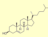
Also at this same one year follow up visit, a repeat cholesterol lab panel showed that Anne’s cholesterol, off the statin drug, had gone up to 330.  The cholesterol had previously been 241 while taking a statin drug.   Above left image: chemical structure of cholesterol courtesy of wikimedia commons

It Has Always Been Elevated

I then asked Anne if her cholesterol had ever been elevated in the past, and she said yes, it has always been elevated in the 300’s her whole life.  Not only that, her parents both have high cholesterol (in the 300’s), and her sisters and brother also have the same thing.  After hearing this, in one of these “Aha moments”, I said to Anne, “you must have familial hypercholesterolemia !”

Statin Drugs for Familial Hypercholesterolemia (FHC)

This complicates the issue.  Indeed, Anne’s mainstream medical doctor has been giving Anne a statin drug for the past 10 years to lower her cholesterol from 330 to 240.  In spite of the “Zero” calcium score, should Anne be taking a statin drug because of the familial hypercholesterolemia (FHC), a risk factor for heart disease ?

Satellite_image_of_Great_Britain_and_Northern_Ireland_in_April_2002Simon Broome Familial Hyperlipidaemia Registry in the UK

The answer comes from a 2008 study from the Simon Broome Familial Hyperlipidaemia Registry in the United Kingdom.(1,2)

Left Image: Satellite Photo of the UK, United Kingdom courtesy of wikimedia commons.

In this study, Three Thousand Four hundred (3400) patients with familial hypercholesterolemia (FHC) were recruited from 21 clinics in the UK and followed for 26 years (46,580 person-years).(1)  What did they find?  Here is a quote from the study:

“Familial hypercholesterolaemia is associated with a substantial excess mortality from coronary heart disease in young adults but may not be associated with a substantial excess mortality in older patients. “(2)

Mortality Depends on Age

The authors looked at data both before and after the availability of statin drugs.

“Before statin drugs were available, mortality from coronary disease was increased nearly 100-fold in young adults with FHC aged 20–39 years, and increased about 4-fold for patients aged 40–59 years, but in those surviving through middle age, risk was similar to the general population.” (1) 

In other words, if a patient survives past 60 with no heart disease, then they are no longer at increased risk.  They have the same risk as the general population.
They went on to say:

Both before and after statins became widely available, there was no excess coronary mortality in patients aged >60 years without known coronary disease. Patients surviving into older age before statins became available were therefore likely to be a highly selected group at lower risk of coronary disease.”(1)

A Highly Selected Group at Lower Risk of Coronary Disease

The Simon Broome Registry of FHC answers our question.(1,2)  At age 61, Anne still has no history of heart disease, and indeed her calcium score of zero indicated no plaque and no heart disease.  Therefore, Anne must be a member of a “highly selected group at lower risk of coronary disease”.

If Anne’s high cholesterol over a lifetime placed her at risk, surely she would have developed some evidence of heart disease by now.  The fact that her calcium score is low means that Anne is at low risk for heart disease.  For Anne, adding a statin drug to prevent heart disease is not necessary and ill advised, since Anne is not at risk.  Statin drugs carry significant adverse effects with reduced memory and cognitive function, muscle pain, neuropathy etc.

An Argument Against the Cholesterol Theory of Heart Disease

Think about it.  Thousands of people with familial hypercholesterolemia who have elevated cholesterol above 330-350 for their entire lives, reach the age of 60 years, and never develop heart disease.  This is one of the strongest arguments against the theory that cholesterol causes heart disease.

Reduce Cholesterol Naturally

The Simon Broome Registry study was re-assuring.  Anne felt comfortable avoiding the statin drugs, and instead use Bergamot, a citrus food supplement called CholestePurePlus.(3,4)  Studies show a 25-30% reduction in cholesterol with Bergamot without adverse side effects associated with statin drugs.(4)

In addition, we plan to follow Anne’s calcium score with an annual Coronary Calcium Score.  Should Anne’s calcium score increase, indicating more plaque formation, then a more aggressive form of intervention would be justified.

Articles with Related Interest

Heart Disease Vitamin C and Linus Pauling

Getting Off Statin Drug Stories

How to Reverse Heart Disease with the Coronary Calcium Score

Cholesterol Lowering Drugs for the Elderly, Bad Idea

Cholesterol Lowering Statin Drugs for Women Just Say No
Jeffrey Dach MD

Links and References

Simon Broome Familial Hyperlipidaemia Register Group  2008

1) http://eurheartj.oxfordjournals.org/content/29/21/2625.full

Eur Heart J. 2008 Nov;29(21):2625-33.
Reductions in all-cause, cancer, and coronary mortality in statin-treated patients with heterozygous familial hypercholesterolaemia: a prospective registry study.
Neil A, Cooper J, Betteridge J, Capps N, McDowell I, Durrington P, Seed M, Humphries SE.Source NIHR School of Primary Care Research, Division Public Health and Primary Health Care, University of Oxford, Old Road Headington, Oxford, UK.

Aims To examine the changes in coronary, all-cause, and cancer mortality in patients with heterozygous familial hypercholesterolaemia (FH) before and after lipid-lowering therapy with statins.
Methods and results A total of 3382 patients (1650 men) aged <80 years were recruited from 21 lipid clinics in the United Kingdom and followed prospectively between 1980 and 2006 for 46 580 person-years. There were 370 deaths, including 190 from coronary heart disease (CHD) and 90 from cancer. The standardized mortality ratio (compared with the population in England and Wales) was calculated before and from 1 January 1992. In patients aged 20–79 years, CHD mortality fell significantly by 37% (95% CI = 7–56) from 3.4- to 2.1-fold excess. Primary prevention resulted in a 48% reduction in CHD mortality from 2.0-fold excess to none, with a smaller reduction of nearly 25% in patients with established disease. Coronary mortality was reduced more in women than in men. In patients without known CHD at registration, all-cause mortality from 1992 was 33% (21–43), lower than in the general population, mainly due to a 37% (21–50) lower risk of fatal cancer.Introduction
Familial hypercholesterolaemia (FH) is an autosomal co-dominant disorder.1 Defects in at least three different genes that code for proteins involved in hepatic clearance of low-density lipoprotein-cholesterol (LDL-C) can cause FH. These include, most commonly, mutations in the gene coding for the LDL-receptor that removes LDL,2 much less commonly in the gene for Apolipoprotein B which is the major protein of the LDL particle, and rarely in the gene coding for an enzyme called PCSK9 involved in degrading the LDL receptor.3 In all cases, this results in an accumulation of LDL in the plasma from birth, and to subsequent development of tendon xanthomas, xanthelasmas, and atheroma.1

In the heterozygous condition, the cumulative risk of a coronary event by the age of 60 years without effective treatment is at least 50% in men and ∼30% in women. Coronary disease occurs ∼10 years earlier in men than in women, with a marked increase in women post-menopausally.4–6

Before effective treatment with HMG-Co reductase inhibitors (statins) became available, mortality from coronary disease was increased nearly 100-fold in young adults aged 20–39 years, and ∼4-fold for patients aged 40–59 years, but in those surviving through middle age risk was similar to the high rates of CHD in the general population of England and Wales.7,8

In most European populations, heterozygous FH affects about one in 500 individuals, but no randomized placebo-controlled clinical outcome trials of statin treatment have been conducted for ethical reasons. Clinical management is therefore largely based on extrapolation from the results of cholesterol-lowering trials conducted in patients with polygenic hypercholesterolaemia;8 from evidence using carotid intima-medial thickness as a surrogate outcome;9 and from a small number of prospective observational studies. The latter include the Simon Broome Register of Familial Hyperlipidaemia, which is a register of patients with heterozygous FH recruited from 21 lipid clinics in Great Britain.7 Earlier results from the register suggested that the prognosis for the heterozygous condition had improved since the introduction and widespread use of statins.10
The aim of this paper was to extend our previous reports7,10–13 by studying an enlarged cohort of 3382 heterozygous patients followed for up to 26 years until the end of 2006, by when the exposure had more than doubled to 46 580 person-years. This has allowed us to examine more informatively the changes in mortality compared with the general population both before and after the routine use of statins.Principal findings  : This large long-term prospective registry study of 3382 patients with heterozygous familial hypercholesterolaemia demonstrates a statistically significant reduction in coronary mortality of about one-third since the widespread use of statins.Primary prevention resulted in a halving in risk of fatal coronary events, with a smaller reduction of nearly one-quarters in patients with established disease. Mortality was reduced more in women than in men for both primary and secondary prevention. Importantly, in patients without known coronary disease at registration, all-cause mortality was significantly lower than in the general population, mainly due to a reduction of more than one-thirds in the risk of fatal cancer. The data also confirm our earlier findings that FH patients are not at a higher risk of fatal stroke.11

However, the Cholesterol Treatment Trialists’ Collaborators systematic prospective meta-analysis,8 which reported the efficacy of cholesterol-lowering treatment using individual patient data for 90 056 participants in 14 randomized trials of statins, found a similar proportional reduction in major coronary events irrespective of age, sex, or previous coronary disease. This differs from the pattern of mortality evident in different sub-groups in our study.

Coronary mortality:  We found that, before and after statins became widely available, there was no excess coronary mortality in patients aged >60 years without known coronary disease at registration

Patients surviving into older age before statins became available were therefore likely to be a highly selected group at lower risk of coronary disease.

All-cause and cancer mortalities: All-cause mortality was significantly reduced by about one-third in patients without known coronary disease at registration, which was mainly due to a reduction in cancer mortality.This is probably attributable to close adherence to advice given as part of routine clinical care to be physically active, make dietary changes,30 avoid obesity,31 and stop smoking.

No increased mortality for men or women after age 60 in familial hypercholesterolemia.

2) http://www.ncbi.nlm.nih.gov/pmc/articles/PMC1671226/?page=
BMJ. 1991 October 12; 303(6807): 893–896.

Risk of fatal coronary heart disease in familial hypercholesterolaemia. Scientific Steering Committee on behalf of the Simon Broome Register Group.

OBJECTIVES–(a) To determine the excess mortality from all causes and from coronary heart disease in patients with familial hypercholesterolaemia; (b) to examine how useful various criteria for selective measurement of cholesterol concentration in cardiovascular screening programmes are in identifying these patients. DESIGN–Prospective cohort study. SETTING–Eleven hospital outpatient lipid clinics in the United Kingdom. PATIENTS–282 men and 244 women aged 20-74 with heterozygous familial hypercholesterolaemia. MAIN OUTCOME MEASURE–Standardised mortality ratio, all adults in England and Wales being taken as standard (standardised mortality ratio = 100 for standard population). RESULTS–The cohort was followed up for 2234 person years during 1980-9. Fifteen of the 24 deaths were due to coronary heart disease, giving a standardised mortality ratio of 386 (95% confidence interval 210 to 639). The excess mortality from this cause was highest at age 20-39 (standardised mortality ratio 9686; 3670 to 21,800) and decreased significantly with age. The standardised mortality ratio for all causes was 183 (117 to 273) and also was highest at age 20-39 (standardised mortality ratio 902; 329 to 1950). There was no significant difference between men and women. Criteria for measurement of cholesterol concentration in cardiovascular screening programmes (family history, presence of myocardial infarction, angina, stroke, corneal arcus, xanthelasma, obesity, hypertension, diabetes, or any of these) were present in 78% of patients. CONCLUSIONS–Familial hypercholesterolaemia is associated with a substantial excess mortality from coronary heart disease in young adults but may not be associated with a substantial excess mortality in older patients.  Criteria for selective measurement of cholesterol concentration in cardiovascular screening programmes identify about three quarters of patients with the clinically overt condition.
——————————

phytosterol Bergamonte™ Citrus Bergamia500 mg.  Risso Polyphenolic Extract (fruit)3) bergamot study   Bergamot Polyphenols The Bergamot orange (Citrus bergamia) is a yellow-colored citrus fruit the size of a lemon mainly grown in the southern Calabria region of Italy. It is distinct from other citrus fruits based on its unique profile and high concentration of flavonoids and flavonoid glycosides. Preliminary cell-based and animal research studies have shown several of these flavonoids to possess anti-atherosclerotic properties, including the ability to inhibit LDL oxidation. Some also have structural similarity to the natural substrate of the HMG-CoA reductase enzyme, which is the major target of statin drugs. Inhibiting this enzyme’s function serves to decrease cholesterol synthesis.

A recent study by Vincenzo Mollace and colleagues in Italy (Fitoterapia, 2011) investigated the effect of bergamot extract high in polyphenols, in both rats and humans. In rats with diet-induced hyperlipidemia, 10 and 20 mg/kg of bergamot polyphenols orally administered daily for 30 days led to significant reductions in total cholesterol, LDL, and triglycerides, with moderate elevations in HDL cholesterol levels seen compared to rats fed the hypercholesterolemic diet alone.

The human study was conducted as a randomized, double-blind, placebo-controlled trial consisting of 237 patients with high cholesterol levels (104 patients with isolated hypercholesterolemia/LDL levels greater than 130 mg/dL) (Group A); 42 patients with elevated cholesterol and triglycerides (Group B); 59 patients with high cholesterol, triglycerides, and blood sugar (Group C); and a final group of 32 patients classified as post–statin therapy who had stopped simvastatin due to musculoskeletal and liver adverse effects (Group D). Each of the above groups was divided into three subgroups. One subgroup received 500 mg of bergamot polyphenols per day; the second received 1000 mg/day; the third group received placebo. The post–statin therapy group received 1500 mg of bergamot polyphenols per day after a 60-day statin washout period. All patients were treated for 30 days.

In aggregate, groups A, B, and C showed an average reduction in total cholesterol of 21.8% and LDL cholesterol of 24.1% and an increase in HDL levels of 22.3% with 500 mg/day. The group taking 1000 mg/day of bergamot polyphenols saw reductions of 29.4% and 36.0% in total cholesterol and LDL, respectively, while achieving an increase in HDL levels of 40.1%. All parameters in the placebo group were essentially unchanged from baseline. In the post-statin group, there was a 25.0% reduction in total cholesterol, 27.6% decrease in LDL levels, and a 23.8% increase in HDL—with none of the side effects seen earlier with statin therapy in this group. Based on these results, the bergamot polyphenols proved to be an effective therapeutic intervention for elevated cholesterol levels as well as a safe and beneficial alternative for individuals intolerant to statin drugs.

4) http://www.ncbi.nlm.nih.gov/pubmed/21056640
Fitoterapia. 2011 Apr;82(3):309-16.

Hypolipemic and hypoglycaemic activity of bergamot polyphenols: from animal models to human studies.Mollace V, Sacco I, Janda E, Malara C, Ventrice D, Colica C, Visalli V, Muscoli S, Ragusa S, Muscoli C, Rotiroti D, Romeo F.
SourceFaculty of Pharmacy, Department of Pharmacobiological Sciences, University Magna Graecia, Catanzaro, Italy.Bergamot juice produces hypolipemic activity in rats though the mechanism remains unclear. Here we investigated on the effect of bergamot extract (BPF) in diet-induced hyperlipemia in Wistar rats and in 237 patients suffering from hyperlipemia either associated or not with hyperglycaemia. BPF, given orally for 30 days to both rats and patients, reduces total and LDL cholesterol levels (an effect accompanied by elevation of cHDL), triglyceride levels and by a significant decrease in blood glucose. Moreover, BPF inhibited HMG-CoA reductase activity and enhanced reactive vasodilation thus representing an efficient phytotherapeutic approach in combating hyperlipemic and hyperglycaemic disorders.

5) Familial hypercholesterolaemia: summary of NICE guidance

6) The benefits of familial hypercholesterolaemia by Uffe Ravnskov  BMJ Rapid Response 4 October 2008

“The NICE guidelines for familial hypercholesterolaemia (FH) are based on the commonly accepted view that early coronary heart disease in FH is caused by high cholesterol.1

Several observations indicate that it is not that simple.

First, studies including only people with FH have shown that both the prevalence and future cardiovascular disease are independent on their blood cholesterol level 2-7 in one of the studies mean cholesterol was even lowest in those who had coronary heart disease (CHD).

In accordance, cholesterol lowering by ileal surpass or by non-statin drugs 9 has no effect in FH, indicating that the small effect obtained with the statins is due to their pleiotropic effects. Most likely, it is their effect on the coagulation system,10-12 as some of the strongest risk factors in FH are high fibrinogen, high factor VIII,13 and high prothrombin,7 because people with FH may have other genetic aberrations as well.7

This interpretation fits well with the fact that atherosclerosis in FH is mainly located to arteries that are exposed to mechanical forces, while premature atherosclerosis is absent in the cerebral arteries, even in homozygous FH.

Even more surprising is that according to the The Simon Broome FH Register Group, the mean life expectancy in FH is as long as for other people; more die from CHD at a young age, but fewer die from cancer and other diseases later in life.

16 These calculations were based on a selection of FH people with close relatives, who had died early, and the authors therefore assumed that the prognosis would have been even better for unselected individuals. Also, before 1900 their life expectancy was longer than for the general population,17 probably because high cholesterol protects against infectious diseases,18 the commonest cause of death at that time.

Therefore, a more appropriate management of FH might be to evaluate the coagulation system and to find appropriate means to correct possible abnormalities. People with FH without such abnormalities should also be told that their high cholesterol is an advantage. The peace of mind following this information should probably be more beneficial for the prevention of CHD than any cholesterol lowering measure.” endquote Uffe RAVNSKOV

References

1. Wierzbicki AS, Humphries SE, Minhas R; Guideline Development Group. Familial hypercholesterolaemia: summary of NICE guidance.BMJ2008;337:a1095

2. Miettinen TA, Gylling H. Mortality and cholesterol metabolism in familial hypercholesterolemia. Long-term follow-up of 96 patients. Arteriosclerosis1988;8:163-7.

3. Hill JS, Hayden MR, Frohlich J, Pritchard PH. Genetic and environmental factors affecting the incidence of coronary artery disease in heterozygous familial hypercholesterolemia. Arterioscler Thromb 1991;11:290-7.

4. Ferrieres J, Lambert J, Lussier-Cacan S, Davignon J. Coronary artery disease in heterozygous familial hypercholesterolemia patients with the same LDL receptor gene mutation. Circulation 1995; 92:290-5.

5. Kroon AA, Ajubi N, van Asten WN, Stalenhoef AF. The prevalence of peripheral vascular disease in familial hypercholesterolaemia J Intern Med1995;238:451-9.

6. Hopkins PN, Stephenson S, Wu LL, Riley WA, Xin Y, Hunt SE. Evaluation of coronary risk factors in patients with heterozygous famlial hypercholesterolema. Am J Cardiol 2001;87:47-553.

7. Jansen AC, van Aalst-Cohen ES, Tanck MW, Cheng S, Fontecha MR, Li J, et al. Genetic determinants of cardiovascular disease risk in familial hypercholesterolemia. Arterioscler Thromb Vasc Biol 2005;25:1475-81.

8. Koivisto PVI, Leinonen H.Peripheral arterial disease in heterozygous familial hypercholesterolemia: no difference between patients with and without partial ileal bypass. Atherosclerosis 1988;70:21-7.

9. Bots ML, Visseren FL, Evans GW, Riley WA, Revkin JH, Tegeler CH, et al. Torcetrapib and carotid intima-media thickness. Lancet 2007;370:153-60.

10. Tremoli E, Folco G, Agradi E, Galli C. Platelet thromboxanes and serum-cholesterol. Lancet 1979;1:107-8.

11. Schrör K. Platelet reactivity and arachidonic acid metabolism in type II hyperlipoproteinaemia and its modification by cholesterol-lowering agents.Eicosanoids 1990; , 67-73.

12. Davì G, Averna M, Catalano I, Barbagallo C, Ganci A, Notarbartolo A, et al. Increased thromboxane biosynthesis in type IIa hypercholesterolemia.Circulation 1992:85:1792-8.

13. Sugrue DD and others. Coronary artery disease and haemostatic variables in heterozygous familial hypercholesterolaemia. Br Heart J1985;53:265-8.

14. Postiglione A, Nappi A, Brunetti A, Soricelli A, Rubba P, Gnasso A, et al. Relative protection from cerebral atherosclerosis of young patients with homozygous familial hypercholesterolemia. Atherosclerosis 1991;90:23-30.

15. Rodriguez G, Bertolini S, Nobili F, Arrigo A, Masturzo P, Elicio N, et al. Regional cerebral blood flow in familial hypercholesterolaemia. Stroke1994;25:831-6.

16. Neil HA, Hawkins MM, Durrington PN, Betteridge DJ, Capps NE, Humphries SE, et al. Non-coronary heart disease mortality and risk of fatal cancer in patients with treated heterozygous familial hypercholesterolaemia: a prospective registry study.Atherosclerosis2005;179:293-7.

17. Sijbrands EJ, Westendorp RG, Defesche JC, de Meier PH, Smelt AH, Kastelein JJ. Mortality over two centuries in large pedigree with familial hypercholesterolaemia: family tree mortality study.BMJ 2001;322:1019-23.

18. Ravnskov U. High cholesterol may protect against infections and atherosclerosis. QJM 2003;96:927-34.

————————————————–++++++++++++++++++++++++++++++

========================================================
http://atvb.ahajournals.org/cgi/content/full/25/7/1475
Arteriosclerosis, Thrombosis, and Vascular Biology. 2005;25:1475.
Atherosclerosis and Lipoproteins Genetic Determinants of Cardiovascular Disease Risk in Familial Hypercholesterolemia

Angelique C.M. Jansen; Emily S. van Aalst-Cohen; Michael W.T. Tanck; Suzanne Cheng; Marcel R. Fontecha; Jia Li; Joep C. Defesche; John J.P. KasteleinFrom the Departments of Vascular Medicine (A.C.M.J., E.S.v.A.-C., J.C.D., J.J.P.K.) and Clinical Epidemiology and Biostatistics (M.W.T.T.), Academic Medical Center, University of Amsterdam, the Netherlands; and the Department of Human Genetics (S.C., M.R.F., J.L.), Roche Molecular Systems, Inc, Alameda, Calif.  Correspondence to John J.P. Kastelein, Department of Vascular Medicine, Academic Medical Center, Meibergdreef 9, P.O.Box 22700, room F4-159.2, 1100 DE Amsterdam, the Netherlands.

Objective— To investigate the contribution of polymorphisms in multiple candidate genes to cardiovascular disease (CVD) risk in a large cohort of patients with heterozygous familial hypercholesterolemia (FH).Methods and Results— We genotyped 1940 FH patients for 65 polymorphisms in 36 candidate genes. During 91.451 person-years, 643 (33.1%) patients had at least 1 cardiovascular event. Multifactorial Cox survival analysis revealed that the G20210A polymorphism in the prothrombin gene was strongly associated with a significantly increased CVD risk (GA versus GG; P<0.001).Conclusions— In a large cohort of FH patients, we found that the G20210A polymorphism in the prothrombin gene is strongly associated with CVD risk. Our results constitute a step forward in the unraveling of the hereditary propensity toward CVD in FH and might lead to better risk stratification and hence to more tailored therapy for CVD prevention.We investigated the contribution of 65 polymorphisms in 36 candidate genes to CVD risk in FH patients and found that the G20210A polymorphism in the prothrombin gene was associated with CVD risk. Our results constitute a step forward in the unraveling of the hereditary propensity toward CVD in FH.

______________________________________

Angiology Volume 60 Number 1 February/March 2009 127-128 # 2009 SAGE Publications. Letter to the Editor.

After the Failure of ENHANCEd Cholesterol Lowering in Familial Hypercholesterolemia, SEAS of Problems with Ezetimibe  by  Luca Mascitelli, MD Uffe Ravnskov, MD, PhD Francesca Pezzetta, MD Ospedale di Tolmezzo Mark R. Goldstein, MD, FACP

Mounting evidence suggests that in familial hypercholesterolemia neither the incidence3 nor the prevalence4 of cardiovascular disease is associated with the lipid levels. This striking observation has been explained by the narrow range of LDL concentrations. 3

The argument is untenable, however, because total and LDL cholesterol in some individuals with familial hypercholesterolemia may be more than twice as high as in others. A possible cause of cardiovascular disease in familial hypercholesterolemia may be inborn errors of the coagulation system.

1 Whayne TF Jr. Is there a problem with ezetimibe or just ENHANCEd hype? Angiology. 2008 Sep 15 [Epub ahead of print].

2. Pedone C, Carbonin P, Kastelein JJ, et al. ENHANCE Investigators. Simvastatin with or without ezetimibe in familial hypercholesterolemia. N Engl J Med. 2008;358: 1431-1443.

3. Jansen AC, van Aalst-Cohen ES, Tanck MW, et al. The contribution of classical risk factors to cardiovascular disease in familial hypercholesterolaemia: data in 2400 patients. J Intern Med. 2004;256:482-490.

4. de Sauvage Nolting PR, Defesche JC, Buirma RJ, Hutten BA, Lansberg PJ, Kastelein JJ. Prevalence and significance of cardiovascular risk factors in a large cohort of patients with familial hypercholesterolaemia. J Intern Med. 2003;253:161-168.

5. SugrueDD, Trayner I,ThompsonGR, et al.Coronary artery disease and haemostatic variables in heterozygous familial hypercholesterolaemia. Br Heart J. 1985;53: 265-268.

6. Jansen AC, van Aalst-Cohen ES, Tanck MW, et al, Genetic determinants of cardiovascular disease risk in familial hypercholesterolemia. Arterioscler Thromb Vasc Biol. 2005;25:1475-1481.

7. Rossebø AB, Pedersen TR, Boman K, et al. for the SEAS Investigators. Intensive lipid lowering with simvastatin and ezetimibe in aortic stenosis. N Engl J Med. 2008;359:1343-1356.

8 Goldstein MR, Mascitelli L, Pezzetta F. The doubleedged sword of statin immunomodulation. Int J Cardiol. 2008 May 15 [Epub ahead of print].

9. Shepherd J, Blauw GJ, Murphy MB, et al, and the PROSPER study group. PROspective Study of Pravastatin in the Elderly at Risk. Pravastatin in elderly individuals at risk of vascular disease (PROSPER): a randomised
controlled trial. Lancet. 2002;360:1623-1630.

10. Bradford PG, Awad AB. Phytosterols as anticancer compounds.
Mol Nutr Food Res. 2007;51:161-170.

11. Ravnskov U. Re: the association between statins and
cancer incidence in a veterans

===================================

http://www.ncbi.nlm.nih.gov/pubmed/15554949
J Intern Med. 2004 Dec;256(6):482-90.

The contribution of classical risk factors to cardiovascular disease in familial hypercholesterolaemia: data in 2400 patients.  Jansen AC, van Aalst-Cohen ES, Tanck MW, Trip MD, Lansberg PJ, Liem AH, van Lennep HW, Sijbrands EJ, Kastelein JJ.  Department of Vascular Medicine, Academic Medical Centre, University of Amsterdam, 1105 AZ Amsterdam, the Netherlands.

OBJECTIVE: To determine the contribution of classical risk factors to the development of cardiovascular disease (CVD) in patients with heterozygous familial hypercholesterolaemia (FH).DESIGN: A retrospective, multi-centre, cohort study. Extensive data were collected by scrutinizing medical records and the use of questionnaires. Multivariate Cox regression was used to study the relationship between potential risk factors and the occurrence of CVD.SETTING AND SUBJECTS: We included 2400 FH patients from 27 Dutch lipid clinics. The diagnosis of FH was based upon the presence of a low-density lipoprotein receptor mutation or upon strict clinical criteria.MAIN OUTCOME MEASURES: Cardiovascular mortality and CVD.RESULTS: During 112.943 person-years, 782 (32.6%) patients had had at least one cardiovascular event. Male gender (RR 2.82, 95% CI 2.37-3.36), smoking (RR 1.67, 95% CI 1.40-1.99), hypertension (RR 1.36, 95% CI 1.06-1.75), diabetes mellitus (RR 2.19, 95% CI 1.36-3.54), low HDL-C (RR 1.37, 95% CI 1.15-1.63) and elevated lipoprotein(a) levels (RR 1.50, 95% CI 1.20-1.79) proved to be independent CVD risk factors. These six risk factors explained 18.7% of the variation in the occurrence of CVD.CONCLUSIONS: Male gender, smoking, hypertension, diabetes mellitus, HDL cholesterol and lipoprotein(a) levels proved to be important risk factors for CVD in FH patients. In addition to the routine institution of statin therapy, controlling these factors needs special attention in the management of this disorder.
———————————–

http://www.ncbi.nlm.nih.gov/pubmed/16141286

Clin Chem. 2005 Nov;51(11):2067-73. Epub 2005 Sep 1.
Lipoprotein(a) is an independent risk factor for cardiovascular disease in heterozygous familial hypercholesterolemia.  Holmes DT, Schick BA, Humphries KH, Frohlich J.  St. Paul’s Hospital Lipid Clinic and the University of British Columbia Department of Pathology and Laboratory Medicine, Vancouver, Canada.Abstract
BACKGROUND: The role of lipoprotein(a) [Lp(a)] as a predictor of cardiovascular disease (CVD) in patients with heterozygous familial hypercholesterolemia (HFH) is unclear. We sought to examine the utility of this lipoprotein as a predictor of CVD outcomes in the HFH population at our lipid clinic.METHODS: This was a retrospective analysis of clinical and laboratory data from a large multiethnic cohort of HFH patients at a single, large lipid clinic in Vancouver, Canada. Three hundred and eighty-eight patients were diagnosed with possible, probable, or definite HFH by strict clinical diagnostic criteria. Multivariate Cox regression analysis was used to study the relationship between several established CVD risk factors, Lp(a), and the age of first hard CVD event.RESULTS: An Lp(a) concentration of 800 units/L (560 mg/L) or higher was a significant independent risk factor for CVD outcomes [hazard ratio (HR) = 2.59; 95% confidence interval (CI), 1.53-4.39; P < 0.001].

Other significant risk factors were male sex [HR = 3.19 (1.79-5.69); P < 0.001] and ratio of total to HDL-cholesterol [1.18 (1.07-1.30); P = 0.001]. A previous history of smoking or hypertension each produced HRs consistent with increased CVD risk [HR = 1.55 (0.92-2.61) and 1.57 (0.90-2.74), respectively], but neither reached statistical significance (both P = 0.10).

LDL-cholesterol was not an independent predictor of CVD risk [HR = 0.85 (0.0.71-1.01); P = 0.07], nor was survival affected by the subcategory of HFH diagnosis (i.e., possible vs probable vs definite HFH).

CONCLUSION: Lp(a) is an independent predictor of CVD risk in a multiethnic HFH population.

——————–

http://www.ncbi.nlm.nih.gov/pubmed/15547022
Heart. 2004 Dec;90(12):1431-7.
Established and emerging coronary risk factors in patients with heterozygous familial hypercholesterolaemia.  Neil HA, Seagroatt V, Betteridge DJ, Cooper MP, Durrington PN, Miller JP, Seed M, Naoumova RP, Thompson GR, Huxley R, Humphries SE. Division of Public Health & Primary Health Care, Institute of Health Sciences, University of Oxford, Old Road, Headington, Oxford OX3 7LF, UK.

Abstract
OBJECTIVES: To assess the clinical and biochemical factors associated with inter-individual variation in susceptibility to coronary artery disease (CAD) in treated heterozygous familial hypercholesterolaemia.

DESIGN: A cross sectional study was conducted of 410 patients recruited from six lipid clinics in the UK.

RESULTS: CAD was documented in 104 of the 211 men and in 55 of the 199 women with mean ages of onset of 43.1 and 46.5 years, respectively. CAD was significantly more common in men (49% v 28%, p < 0.001) and in patients who had smoked cigarettes versus patients who had never smoked (51% v 28%, p < 0.001). After adjusting for age, sex, and current smoking status, there were no significant differences between patients with or without CAD in lipoprotein(a), homocysteine, fibrinogen, plasminogen activator inhibitor-1, white blood cell count, body mass index, glucose, triglyceride or total cholesterol. However, high density lipoprotein (HDL) cholesterol concentrations were significantly lower in those with CAD (6%, 95% confidence interval (CI) 1% to 11%, p = 0.03) and this difference was greater in women than men (12% v 2%, p = 0.041).

CONCLUSIONS: These results indicate that emerging coronary risk factors appear not to be associated with CAD in adults with treated familial hypercholesterolaemia, but the strong association with smoking suggests that patients should be identified early in childhood and discouraged from ever starting to smoke.

———————-

http://www.ncbi.nlm.nih.gov/pmc/articles/PMC2563208/
J Med Genet. 2006 December; 43(12): 943–949.

Genetic causes of familial hypercholesterolaemia in patients in the UK: relation to plasma lipid levels and coronary heart disease risk.  S E Humphries, R A Whittall, C S Hubbart, S Maplebeck, J A Cooper, A K Soutar, R Naoumova, G R Thompson, M Seed, P N Durrington, J P Miller, D J B Betteridge, and H A W Neil, for the Simon Broome Familial Hyperlipidaemia Register Group and Scientific Steering Committee  S E Humphries, R A Whittall, C S Hubbart, S Maplebeck, J A Cooper, Centre for Cardiovascular Genetics, British Heart Foundation Laboratories, Royal Free and University College London Medical School, London, UKA K Soutar, R Naoumova, G R Thompson, Medical Research Council Clinical Sciences Centre, Imperial College Faculty of Medicine, Hammersmith Hospital, London, UK

—————————————-

http://www.atherosclerosis-journal.com/article/S0021-9150(04)00544-1/abstract
Atherosclerosis Volume 179, Issue 2 , Pages 293-297, April 2005

Non-coronary heart disease mortality and risk of fatal cancer in patients with treated heterozygous familial hypercholesterolaemia: a prospective registry study
H.A.W. Neil et al.

The prognosis from coronary heart disease (CHD) for patients with heterozygous familial hypercholesterolaemia has improved substantially since the introduction of HMG Co-A reductase inhibitors (statins), but the effect of lipid-lowering drug therapy combined with dietary and life style advice on non-coronary mortality and the risk of fatal cancer is unclear.

Methods:The cohort of 2871 patients was recruited from 21 outpatient lipid clinics in the UK from 1980 to 1998 and was followed for 22,992 person-years. The standardised mortality ratio (SMR) was calculated from the ratio of the number of deaths observed to the number expected in the general population of England and Wales.

Results:There were 169 deaths, including 102 (60.4%) from CHD, and 32 (18.9%) from cancer. The SMR for CHD was 2.5-fold higher than in the general population (95% CI 2.1, 3.1), but the all-cause SMR was not increased (1.1, 95% CI 0.9, 1.3) and non-coronary mortality was significantly lower in men (0.5, 95% CI 0.3, 0.7) and women (0.6, 95% CI 0.4, 0.9). The SMR for all cancers was significantly reduced (0.6, 95% CI 0.4, 0.8) with an 80% reduction in fatal cancers of the respiratory and intra-thoracic organs and a non-significant reduction in fatal cancers of the genitourinary and digestive organs.

Conclusions:Although the study cannot exclude the possibility that statins have anti-cancer activity, the results strongly suggest that giving advice to consume a healthy diet, increase physical activity and stop smoking is associated with a substantial reduction in mortality from cancer.

——————————————————-
http://www.ncbi.nlm.nih.gov/pmc/articles/PMC31037/?tool=pubmed

BMJ. 2001 April 28; 322(7293): 1019–1023.

Mortality over two centuries in large pedigree with familial hypercholesterolaemia: family tree mortality study.Sijbrands EJ, Westendorp RG, Defesche JC, de Meier PH, Smelt AH, Kastelein JJ.  Department of Vascular Medicine and General Internal Medicine, Academic Medical Centre, Meibergdreef 9, 1105 AZ Amsterdam, Netherlands.

To estimate all cause mortality from untreated familial hypercholesterolaemia free from selection for coronary artery disease.

Design  Family tree mortality study.Setting  Large pedigree in Netherlands traced back to a single pair of ancestors in the 19th century.Subjects  All members of pedigree aged over 20 years with 0.5 probability of carrying a mutation for familial hypercholesterolaemia.Main outcome measure  All cause mortality.

Results A total of 70 deaths took place among 250 people analysed for 6950 person years. Mortality was not increased in carriers of the mutation during the 19th and early 20th century; it rose after 1915, reached its maximum between 1935 and 1964 (standardised mortality ratio 1.78, 95% confidence interval 1.13 to 2.76; P=0.003), and fell thereafter. Mortality differed significantly between two branches of the pedigree (relative risk 3.26, 95% confidence interval 1.74 to 6.11; P=0.001).

Conclusions  Risk of death varies significantly among patients with familial hypercholesterolaemia. This large variability over time and between branches of the pedigree points to a strong interaction with environmental factors. Future research is required to identify patients with familial hypercholesterolaemia who are at extreme risk and need early and vigorous preventive measures.

———————————————

http://www.ncbi.nlm.nih.gov/pmc/articles/PMC2583391/?tool=pubmed
http://www.ncbi.nlm.nih.gov/pubmed/19001495   free full
BMJ. 2008 Nov 11;337:a2423.
Efficacy of statins in familial hypercholesterolaemia: a long term cohort study.
Versmissen J, Oosterveer DM, Yazdanpanah M, Defesche JC, Basart DC, Liem AH, Heeringa J, Witteman JC, Lansberg PJ, Kastelein JJ, Sijbrands EJ.
SourceDepartment of Internal Medicine, Erasmus University Medical Centre, PO box 2040, 3000 CA Rotterdam, Netherlands.

Abstract
OBJECTIVE: To determine the efficacy of statin treatment on risk of coronary heart disease in patients with familial hypercholesterolaemia.

DESIGN: Cohort study with a mean follow-up of 8.5 years.
SETTING: 27 outpatient lipid clinics.
SUBJECTS: 2146 patients with familial hypercholesterolaemia without prevalent coronary heart disease before 1 January 1990.
MAIN OUTCOME MEASURES: Risk of coronary heart disease in treated and “untreated” (delay in starting statin treatment) patients compared with a Cox regression model in which statin use was a time dependent variable.

RESULTS: In January 1990, 413 (21%) of the patients had started statin treatment, and during follow-up another 1294 patients (66%) started after a mean delay of 4.3 years. Most patients received simvastatin (n=1167, 33 mg daily) or atorvastatin (n=211, 49 mg daily). We observed an overall risk reduction of 76% (hazard ratio 0.24 (95% confidence interval 0.18 to 0.30), P<0.001). In fact, the risk of myocardial infarction in these statin treated patients was not significantly greater than that in an age-matched sample from the general population (hazard ration 1.44 (0.80 to 2.60), P=0.23).

CONCLUSION: Lower statin doses than those currently advised reduced the risk of coronary heart disease to a greater extent than anticipated in patients with familial hypercholesterolaemia. With statin treatment, such patients no longer have a risk of myocardial infarction significantly different from that of the general population.
We finally compared the risk of myocardial infarction in patients with familial hypercholesterolaemia who were older than 55 years (n=261, 64 men) with that in 1975 people in a subgroup of the participants in the Rotterdam study. The mean age in both subgroups was 61.6 years and both had 24.5% men as a result of stratified selection from the Rotterdam study. The absolute risk of myocardial infarction was 6.7/1000 person years in our statin treated patients, 60.5/1000 person years in our untreated patients, and 4.1/1000 person years in the sample from the Rotterdam study. Event-free survival of our statin treated patients was not significantly different from that of the Rotterdam study sample (log rank test P=0.07), whereas our untreated patients clearly had a higher risk of coronary heart disease (log rank test P<0.001) (fig 4​4).). After adjustment for year of birth and sex, the point estimate of risk of myocardial infarction in our treated patients with familial hypercholesterolaemia was higher than the risk in the subgroup of the Rotterdam study, but this was not significant (hazard ratio 1.44 (0.80 to 2.60), P=0.23), whereas the risk in our untreated patients was 8.7 times higher (hazard ratio 8.69 (4.77 to 15.82), P<0.001).

We report here that relatively modest doses of statins reduced the risk of coronary heart disease by about 80% in patients with familial hypercholesterolaemia. This is a much more pronounced reduction than was anticipated based on secular trends in earlier studies.7 8 We also observed that statin treated patients older than 55 years had a risk of myocardial infarction approaching that of the general population. Finally, men and women experienced similar risk reductions in our study.

illustration showing normal mortality curve for over 55 w/ fam hyper
http://www.ncbi.nlm.nih.gov/pmc/articles/PMC2583391/figure/fig4/

———————-

Familial hypercholesterolemia

———————————–

Cholesterol Freatment Trialists’ (CTT) Collaborators.  2005

http://www.ncbi.nlm.nih.gov/pubmed/16214597?dopt=Abstract
Cholesterol Freatment Trialists’ (CTT) Collaborators. Efficacy and safety of cholesterol-lowering treatment: prospective meta-analysis of data from 90,056 participants in 14 randomised trials of statins. Lancet 2005;366:1267-1278.
Lancet. 2005 Oct 8;366(9493):1267-78. .
Efficacy and safety of cholesterol-lowering treatment: prospective meta-analysis of data from 90,056 participants in 14 randomised trials of statins.
Baigent C, Keech A, Kearney PM, Blackwell L, Buck G, Pollicino C, Kirby A, Sourjina T, Peto R, Collins R, Simes R; Cholesterol Treatment Trialists’ (CTT) Collaborators.

Results of previous randomised trials have shown that interventions that lower LDL cholesterol concentrations can significantly reduce the incidence of coronary heart disease (CHD) and other major vascular events in a wide range of individuals. But each separate trial has limited power to assess particular outcomes or particular categories of participant.
METHODS:A prospective meta-analysis of data from 90,056 individuals in 14 randomised trials of statins was done. Weighted estimates were obtained of effects on different clinical outcomes per 1.0 mmol/L reduction in LDL cholesterol.
FINDINGS:During a mean of 5 years, there were 8186 deaths, 14,348 individuals had major vascular events, and 5103 developed cancer. Mean LDL cholesterol differences at 1 year ranged from 0.35 mmol/L to 1.77 mmol/L (mean 1.09) in these trials. There was a 12% proportional reduction in all-cause mortality per mmol/L reduction in LDL cholesterol (rate ratio [RR] 0.88, 95% CI 0.84-0.91; p<0.0001). This reflected a 19% reduction in coronary mortality (0.81, 0.76-0.85; p<0.0001), and non-significant reductions in non-coronary vascular mortality (0.93, 0.83-1.03; p=0.2) and non-vascular mortality (0.95, 0.90-1.01; p=0.1). There were corresponding reductions in myocardial infarction or coronary death (0.77, 0.74-0.80; p<0.0001), in the need for coronary revascularisation (0.76, 0.73-0.80; p<0.0001), in fatal or non-fatal stroke (0.83, 0.78-0.88; p<0.0001), and, combining these, of 21% in any such major vascular event (0.79, 0.77-0.81; p<0.0001). The proportional reduction in major vascular events differed significantly (p<0.0001) according to the absolute reduction in LDL cholesterol achieved, but not otherwise. These benefits were significant within the first year, but were greater in subsequent years. Taking all years together, the overall reduction of about one fifth per mmol/L LDL cholesterol reduction translated into 48 (95% CI 39-57) fewer participants having major vascular events per 1000 among those with pre-existing CHD at baseline, compared with 25 (19-31) per 1000 among participants with no such history. There was no evidence that statins increased the incidence of cancer overall (1.00, 0.95-1.06; p=0.9) or at any particular site.
INTERPRETATION:  Statin therapy can safely reduce the 5-year incidence of major coronary events, coronary revascularisation, and stroke by about one fifth per mmol/L reduction in LDL cholesterol, largely irrespective of the initial lipid profile or other presenting characteristics. The absolute benefit relates chiefly to an individual’s absolute risk of such events and to the absolute reduction in LDL cholesterol achieved. These findings reinforce the need to consider prolonged statin treatment with substantial LDL cholesterol reductions in all patients at high risk of any type of major vascular event.

Mohrschladt 2004

http://www.ncbi.nlm.nih.gov/pubmed/15019543/
Atherosclerosis. 2004 Feb;172(2):329-35.
Cardiovascular disease and mortality in statin-treated patients with familial hypercholesterolemia.  Mohrschladt MF, Westendorp RG, Gevers Leuven JA, Smelt AH.
Source  Department of General Internal Medicine, Leiden University Medical Center, P.O. Box 9600, 2300 RC Leiden, The Netherlands.

Patients with familial hypercholesterolemia (FH) are at an increased risk of premature cardiovascular disease (CVD). The benefits of statin therapy are not well known since no placebo controlled studies have been performed in these patients. The aim of this study was to determine the CVD event and mortality risk in statin-treated patients with FH. A total of 345 FH patients were followed prospectively for 8 years. Mortality from CVD was compared to that of the general population. The absolute risk of CVD in patients without a previous history of CVD was 3% per year for men and 1.6% for women. Mortality from CVD in patients without a previous history was 1.4-fold (95% CI = 0.6-3.3) increased and ischaemic heart disease (IHD) mortality was 2.6-fold (95% CI = 1.1-6.3) higher compared to the general population.

This mortality risk was highest in patients aged 40-59 years.

Female FH patients had no increased CVD or IHD mortality risk. Over a period of 8 years the event risk of patients with a history of CVD was almost 30% per year under age 40 years and 15% in patients aged 60 years and over. When compared to the general population, mortality from other causes than CVD was lower for patients with FH, the relative risks not reaching statistical significance. The relative risk of mortality from all causes was 1.5 (P < 0.05) for men and 1.0 for women. In conclusion, male patients with FH, treated from middle-age with statins remain at an increased risk of developing CVD.

http://www.ncbi.nlm.nih.gov/pubmed/21565855
Heart. 2011 Jul;97(14):1151-7. doi: 10.1136/hrt.2010.220699. Epub 2011 May 12.
CT coronary plaque burden in asymptomatic patients with familial hypercholesterolaemia.
Neefjes LA, Ten Kate GJ, Rossi A, Galema-Boers AJ, Langendonk JG, Weustink AC, Moelker A, Nieman K, Mollet NR, Krestin GP, Sijbrands EJ, de Feyter PJ.
SourceDepartment of Radiology and Cardiology, Erasmus Medical Center Rotterdam, CA, Rotterdam, The Netherlands.OBJECTIVE:To determine the calcium score and coronary plaque burden in asymptomatic statin-treated patients with heterozygous familial hypercholesterolaemia (FH) compared with a control group of patients with low probability of coronary artery disease, having non-anginal chest pain, using CT.
DESIGN, SETTING AND PATIENTS:101 asymptomatic patients with FH (mean age 53 ± 7 years; 62 men) and 126 patients with non-anginal chest pain (mean age 56 ± 7 years; 80 men) underwent CT calcium scoring and CT coronary angiography. All patients with FH were treated with statins during a period of 10 ± 8 years before CT. The coronary calcium score and plaque burden were determined and compared between the two patient groups.
RESULTS:The median total calcium score was significantly higher in patients with FH (Agatston score = 87, IQR 5-367) than in patients with non-anginal chest pain (Agatston score = 7, IQR 0-125; p < 0.001). The overall coronary plaque burden was significantly higher in patients with FH (p < 0.01). Male patients with FH, whose low-density lipoprotein cholesterol levels were reduced by statins below 3.0 mmol/l, had significantly less coronary calcium (p < 0.01) and plaque burden (p = 0.02).
CONCLUSION:The coronary plaque burden is high in asymptomatic middle-aged patients with FH despite intense statin treatment.

http://www.ncbi.nlm.nih.gov/pubmed/22460051
Atherosclerosis. 2012 Jun;222(2):468-72. Detection of subclinical atherosclerosis in familial hypercholesterolemia using non-invasive imaging modalities. Caballero P, Alonso R, Rosado P, Mata N, Fernández-Friera L, Jiménez-Borreguero LJ, Badimon L, Mata P.
Source Department of Radiology, Hospital de la Princesa, Madrid, Spain.

To investigate the extent of subclinical atherosclerosis in asymptomatic familial hypercholesterolemia (FH) patients using non-invasive images techniques.
PATIENTS, METHODS AND RESULTS:
The atherosclerotic burden of 36 molecularly defined FH patients (18 males, 45.7±10.9 years) without evidence of cardiovascular disease receiving lipid-lowering treatment and 19 (47.8±11.3 years) controls was investigated. Descending thoracic aorta magnetic resonance imaging (MRI) was performed in a 1.5 T equipment with T1 and T2 sequences to characterize atherosclerotic plaques and to measure aortic wall volumen. Carotid intima-media thickness (cIMT) and presence of plaques were measured using B-mode carotid ultrasound. Mean aortic wall volumen, cIMT and atherosclerotic plaques in aorta were significantly higher in FH cases (P<0.001). A significant correlation between aortic wall volume and cIMT was observed (P<0.01). Aortic MRI detected plaques in 94% and carotid ultrasound in 14% of cases. Lipid-rich plaques were observed only in FH cases (33%) and were associated with family history of premature coronary artery disease (P<0.05).
CONCLUSIONS:Asymptomatic middle-aged FH patients have significantly higher atherosclerotic burden than controls. cIMT has shown a significant correlation with aortic wall volume and MRI allowed the detection of lipid-rich plaques in FH subjects that were associated with family history of premature coronary artery disease.

http://www.ncbi.nlm.nih.gov/pubmed/22018443
Atherosclerosis. 2011 Dec;219(2):721-7.Accelerated subclinical coronary atherosclerosis in patients with familial hypercholesterolemia.
Neefjes LA, Ten Kate GJ, Alexia R, Nieman K, Galema-Boers AJ, Langendonk JG, Weustink AC, Mollet NR, Sijbrands EJ, Krestin GP, de Feyter PJ.
Source Department of Radiology, Erasmus Medical Center, Rotterdam, The Netherlands.

We determined the extent, severity, distribution and type of coronary plaques in cardiac asymptomatic patients with familial hypercholesterolemia (FH) using computed tomography (CT).
BACKGROUND:FH patients have accelerated progression of coronary artery disease (CAD) with earlier major adverse cardiac events. Non-invasive CT coronary angiography (CTCA) allows assessing the coronary plaque burden in asymptomatic patients with FH.
MATERIALS AND METHODS:A total of 140 asymptomatic statin treated FH patients (90 men; mean age 52 ± 8 years) underwent CT calcium scoring (Agatston) and CTCA using a Dual Source CT scanner with a clinical follow-up of 29 ± 8 months. The extent, severity (obstructive or non-obstructive plaque based on >50% or <50% lumen diameter reduction), distribution and type (calcified, non-calcified, or mixed) of coronary plaque were evaluated.
RESULTS:The calcium score was 0 in 28 (21%) of the patients. In 16% of the patients there was no CT-evidence of any CAD while 24% had obstructive disease. In total 775 plaques were detected with CT coronary angiography, of which 11% were obstructive. Fifty four percent of all plaques were calcified, 25% non-calcified and 21% mixed. The CAD extent was related to gender, treated HDL-cholesterol and treated LDL-cholesterol levels. There was a low incidence of cardiac events and no cardiac death occurred during follow-up.
CONCLUSION:Development of CAD is accelerated in intensively treated male and female FH patients. The extent of CAD is related to gender and cholesterol levels and ranges from absence of plaque in one out of 6 patients to extensive CAD with plaque causing >50% lumen obstruction in almost a quarter of patients with FH.

http://www.ncbi.nlm.nih.gov/pubmed/20980000
Atherosclerosis. 2010 Dec;213(2):486-91. Evaluation of subclinical atherosclerosis by computed tomography coronary angiography and its association with risk factors in familial hypercholesterolemia.
Miname MH, Ribeiro MS 2nd, Parga Filho J, Avila LF, Bortolotto LA, Martinez LR, Rochitte CE, Santos RD.
Source Lipid Clinic Heart Institute (InCor), University of São Paulo, Medical School Hospital, São Paulo, Brazil.

Increasing age and cholesterol levels, male gender, and family history of early coronary heart disease (CHD) are associated with early onset of CHD in familial hypercholesterolemia (FH).
OBJECTIVE:Assess subclinical atherosclerosis by computed tomography coronary angiography (CTCA) and its association with clinical and laboratorial parameters in asymptomatic FH subjects.
METHODS:102 FH subjects (36% male, 45 ± 13 years, LDL-c 280 ± 54 mg/dL) and 35 controls (40% male, 46 ± 12 years, LDL-c 103 ± 18 mg/dL) were submitted to CTCA. Plaques were divided into calcified, mixed and non-calcified; luminal stenosis was characterized as >50% obstruction.
RESULTS:FH had a greater atherosclerotic burden represented by higher number of patients with: plaques (48% vs. 14%, p=0.0005), stenosis (19% vs. 3%, p=0.015), segments with plaques (2.05 ± 2.85 vs.0.43 ± 1.33, p=0.0016) and calcium scores (55 ± 129 vs. 38 ± 140, p=0.0028). After multivariate analysis, determinants of plaque presence were increasing age (OR=2.06, for age change of 10 years, CI95%: 1.38-3.07, p<0.001) and total cholesterol (OR=1.86, for cholesterol change by 1 standard deviation, CI95%: 1.09-3.15, p=0.027). Coronary calcium score was associated with the presence of stenosis (OR=1.54; CI95%: 1.27-1.86, p<0.001, for doubling the calcium score). Male gender was directly associated with the presence of non-calcified plaques (OR: 15.45, CI95% 1.72-138.23, p=0.014) and inversely with calcified plaques (OR=0.21, CI95%: 0.05-0.84, p=0.027). Family history of early CHD was associated with the presence of mixed plaques (OR=4.90, CI95%: 1.32-18.21, p=0.018).
CONCLUSIONS: Patients with FH had an increased burden of coronary atherosclerosis by CTCA. The burden of atherosclerosis and individual plaque subtypes differed with the presence of other associated risk factors, with age and cholesterol being most important. A coronary calcium score of zero ruled out obstructive disease in this higher risk population.

http://www.ncbi.nlm.nih.gov/pubmed/23340035
Am J Cardiol. 2013 Apr 1;111(7):955-61.
Coronary computed tomographic angiographic findings in asymptomatic patients with heterozygous familial hypercholesterolemia and null allele low-density lipoprotein receptor mutations.
Viladés Medel D, Leta Petracca R, Carreras Costa F, Cardona Olle M, Barros Membrilla A, Hidalgo Perez JA, Pujadas Olano S, Alomar Serrallach X, Franco Peral M, Pons-Lladó G.
Source Cardiac Imaging Unit, Cardiology Department, Universitat Autònoma de Barcelona, Barcelona, Spain.

Heterozygous familial hypercholesterolemia (HeFH) can be associated with early coronary artery disease (CAD) in asymptomatic patients. The objectives of the present study were to assess the prevalence and magnitude of subclinical CAD in patients with HeFH using coronary computed tomographic angiography (CCTA) and to determine the clinical and genetic profile of those at the greatest risk of CAD. The study included 50 consecutive patients with HeFH diagnosed according to the Dutch Lipid Clinic Network criteria and a control group of 70 healthy subjects. The findings from CCTA for the patients with HeFH were compared with those from the control group, who had been referred for CCTA as a part of a preventive medical examination. In 82% of the patients with HeFH, genetic DNA was screened for low-density lipoprotein receptor (LDLR) gene mutations using a microarray. CCTA revealed a significantly greater Agatston calcium score in the study group than in the control group (260 vs 46; p = 0.002). The prevalence of CAD in the patients with HeFH was 48%. It was significant in 26%, involving mainly the proximal segments of the coronary arteries. In the control group, the prevalence of CAD was 33% and was significant in 5% (p <0.05 for prevalence and severity of CAD compared to patients with HeFH). In those with HeFH, increased age, null allele LDLR mutations, and low high-density lipoprotein blood levels at diagnosis showed a statistically significant association with CAD (p <0.05). In conclusion, patients with HeFH present with a greater prevalence, extension, and severity of subclinical CAD than the general population. Increased age, low high-density lipoprotein levels, and LDLR null allele mutations are related to the occurrence of CAD. CCTA has emerged as a useful technique for the screening of subclinical CAD in patients with HeFH.
Evidence that men with familial hypercholesterolemia can avoid early coronary death. An analysis of 77 gene carriers in four Utah pedigrees.
Williams RR, Hasstedt SJ, Wilson DE, Ash KO, Yanowitz FF, Reiber GE, Kuida H.  JAMA. 1986 Jan 10;255(2):219-24.

Abstract  To study the genetic influence on serum cholesterol levels and early coronary heart disease, 1,134 individuals were screened from 18 Utah pedigrees. In most pedigrees, serum cholesterol appeared to be a purely polygenic trait, with 54% heritability. In four pedigrees with dominant familial hypercholesterolemia, male heterozygotes had a mean serum cholesterol level of 352 mg/dL, myocardial infarction at an average age of 42 years, and coronary death at an average age of 45 years. An informative pedigree structure allowed the identification of four ancestral males born before 1880 who carried this lethal gene and survived to ages 62, 68, 72, and 81 years. This suggests that some healthy life-style factors protected these men against the expression of a gene that has led to coronary disease by age 45 years in all of their heterozygous great-grandsons. One heterozygote showed a drop in serum cholesterol level from 426 to 248 mg/dL, with strict adherence to a low-fat diet without drugs. These observations should help encourage physicians to try harder to identify and help such individuals.

http://circ.ahajournals.org/content/92/3/290.long
http://www.ncbi.nlm.nih.gov/pubmed/7634440
Circulation. 1995 Aug 1;92(3):290-5.
Coronary artery disease in heterozygous familial hypercholesterolemia patients with the same LDL receptor gene mutation.
Ferrières J, Lambert J, Lussier-Cacan S, Davignon J.
SourceDépartement de médecine sociale et préventive, Faculté de Médecine, Université de Montréal, Quebec, Canada.
Abstract
BACKGROUND:Familial hypercholesterolemia (FH), an autosomal codominant disease, is characterized by high levels of LDL cholesterol and a high incidence of coronary artery disease (CAD). To date, genetic heterogeneity has hindered the proper assessment of the relation between risk factors and CAD in FH patients.

METHODS AND RESULTS:  We studied the association between CAD and common risk factors in a sample of 263 French Canadian FH patients (147 women, 116 men) carrying the same > 10-kb deletion of the LDL receptor gene. Thirty-five women and 54 men had CAD. The mean age of onset of CAD was 45.6 +/- 12.7 years in women and 38.8 +/- 9.4 years in men. Multiple logistic regression analyses were performed to test the association between CAD and age, tendon xanthomas, cigarette smoking, hypertension, diabetes mellitus, apolipoprotein E polymorphism, total plasma cholesterol, triglycerides, VLDL cholesterol, LDL cholesterol, HDL cholesterol, and lipoprotein(a) [Lp(a)]. In FH women, significant multivariate predictors were age (odds ratio, 1.10 for 1 year; P < .0001), VLDL cholesterol (odds ratio, 3.85 for 1 natural log unit; P < .002), and LDL cholesterol (odds ratio, 1.42 for 1 mmol/L; P < .02). In FH men, age (odds ratio, 1.08 for 1 year; P < .0001) and HDL cholesterol (odds ratio, 0.14 for 1 mmol/L; P = .05) were significant predictors of disease. Lp(a) was not a significant predictor in univariate or multivariate analyses.
CONCLUSIONS:This study suggests that increased risk of CAD in FH is not solely due to elevated LDL cholesterol levels and demonstrates a sex-specific lipoprotein influence on CAD in a large sample of FH patients carrying the same LDL receptor gene defect.

http://atvb.ahajournals.org/content/25/7/1475.long
http://www.ncbi.nlm.nih.gov/pubmed/15879303
Arterioscler Thromb Vasc Biol. 2005 Jul;25(7):1475-81. Epub 2005 May 5.
Genetic determinants of cardiovascular disease risk in familial hypercholesterolemia.
Jansen AC, van Aalst-Cohen ES, Tanck MW, Cheng S, Fontecha MR, Li J, Defesche JC, Kastelein JJ.
Source  Department of Vascular Medicine, Academic Medical Center, Amsterdam, The Netherlands.
Abstract OBJECTIVE:  To investigate the contribution of polymorphisms in multiple candidate genes to cardiovascular disease (CVD) risk in a large cohort of patients with heterozygous familial hypercholesterolemia (FH).
METHODS AND RESULTS:  We genotyped 1940 FH patients for 65 polymorphisms in 36 candidate genes. During 91.451 person-years, 643 (33.1%) patients had at least 1 cardiovascular event. Multifactorial Cox survival analysis revealed that the G20210A polymorphism in the prothrombin gene was strongly associated with a significantly increased CVD risk (GA versus GG; P<0.001).
CONCLUSIONS:  In a large cohort of FH patients, we found that the G20210A polymorphism in the prothrombin gene is strongly associated with CVD risk. Our results constitute a step forward in the unraveling of the hereditary propensity toward CVD in FH and might lead to better risk stratification and hence to more tailored therapy for CVD prevention.

http://www.ncbi.nlm.nih.gov/pmc/articles/PMC481754/
Br Heart J. 1985 Mar;53(3):265-8.
Coronary artery disease and haemostatic variables in heterozygous familial hypercholesterolaemia.  Sugrue DD, Trayner I, Thompson GR, Vere VJ, Dimeson J, Stirling Y, Meade TW.

Abstract  Haemostatic variables were measured in 61 patients with heterozygous familial hypercholesterolaemia, 32 of whom had evidence of coronary heart disease. Age adjusted mean concentrations of plasma fibrinogen and factor VIII were significantly higher in these patients than in the 29 patients without coronary heart disease, but there were no significant differences in serum lipid concentrations between the two groups. Comparisons in 30 patients taking and not taking lipid lowering drugs showed lower values for low density lipoprotein cholesterol, high density lipoprotein cholesterol and antithrombin III, and a higher high density lipoprotein ratio while receiving treatment. The results suggest that hypercoagulability may play a role in the pathogenesis of coronary heart disease in patients with familial hypercholesterolaemia.

http://www.ncbi.nlm.nih.gov/pubmed/9920511
Atherosclerosis. 1999 Jan;142(1):105-12.
Mortality in treated heterozygous familial hypercholesterolaemia: implications for clinical management. Scientific Steering Committee on behalf of the Simon Broome Register Group.  [No authors listed]

Abstract Clinical management of heterozygous familial hypercholesterolaemia is largely based on evidence from a small number of observational studies and extrapolation from the results of clinical trials of lipid-lowering in patients with polygenic hypercholesterolaemia The objectives of this study were (i) to determine the absolute and relative mortality of patients with treated heterozygous familial hypercholesterolaemia, (ii) to estimate the effect of changes in treatment efficacy on mortality trends over time, and (iii) to examine the implications of these findings for patient management. A cohort of 605 men and 580 women aged 20-79 years with heterozyous familial hypercholesterolaemia were recruited from 21 out-patient lipid clinics in the UK. Patients were followed prospectively from 1980 to 1995 for 8770 person-years. Absolute mortality was calculated, and relative risk was expressed as the ratio of the number of observed deaths to the number expected in the general population of England and Wales. Forty six of the 73 deaths were due to coronary heart disease. In women aged 20-39, despite treatment, the relative risk of a fatal coronary event was increased 125-fold (95% confidence intervals 15-451) and the annual coronary mortality was 0.17%. In men aged 20-39 the relative risk was increased 48-fold (17-105) and the annual coronary mortality was 0.46%. The relative risk decreased with age but the absolute risk increased. For men and women aged 60-79, the annual coronary mortality was 1.1% representing a significant excess mortality for women (relative risk 2.6, 1.3-4.5) but not for men (RR 1.1, 0.5-2.3). Non-coronary mortality was not increased at any age (RR for all ages 0.68, 0.45-0.99). There was a decline in the relative risk for coronary mortality in patients aged 20-59 from an eight-fold (4.8-7.2) increased risk before 1992 to 3.7 (1.6-7.2.) thereafter (P=0.08). The results suggest that the prognosis for patients with heterozygous familial hypercholesterolaemia has improved with the introduction of more effective treatment, and that lipid-lowering therapy is not associated with increased non-coronary mortality. These findings and the excess coronary mortality observed suggest that all affected adult men and post-menopausal women should be treated with HMG-CoA reductase inhibitors.Benefits of high cholesterol

http://www.ravnskov.nu/the%20benefits%20of%20high%20C.htm

http://www.ncbi.nlm.nih.gov/pubmed/20952373
Age Ageing. 2010 Nov;39(6):674-80. Lipid-lowering treatment to the end? A review of observational studies and RCTs on cholesterol and mortality in 80+-year olds.  Petersen LK, Christensen K, Kragstrup J.
Source  Research Unit of Epidemiology, Danish Aging Research Center, University of Southern Denmark, Odense, Denmark.

People aged 80 or older are the fastest growing population in high-income countries. One of the most common causes of death among the elderly is the cardiovascular disease (CVD). Lipid-lowering treatment is common, e.g. one-third of 75-84-year-old Swedes are treated with statins. The assumption that hypercholesterolaemia is a risk factor at the highest ages seems to be based on extrapolation from younger adults. A review of observational studies shows a trend where all-cause mortality was highest when total cholesterol (TC) was lowest (‘a reverse J-shaped’ association between TC and all-cause mortality). Low TC (<5.5 mmol/l) is associated with the highest mortality rate in 80+-year olds. No clear optimal level of TC was identified. A review of the few randomised controlled trials including 80+-year olds did not provide evidence of an effect of lipid-lowering treatment on total mortality in 80+-year-old people. There is not sufficient data to recommend anything regarding initiation or continuation of lipid-lowering treatment for the population aged 80+, with known CVD, and it is even possible that statins may increase all-cause mortality in this group of elderly individuals without CVD.

————————-

Jeffrey Dach MD
http://www.drdach.com/
http://www.naturalmedicine101.com/
http://www.truemedmd.com/

Disclaimer click here: http://www.drdach.com/wst_page20.html

The reader is advised to discuss the comments on these pages with his/her personal physicians and to only act upon the advice of his/her personal physician. Also note that concerning an answer which appears as an electronically posted question, I am NOT creating a physician — patient relationship. Although identities will remain confidential as much as possible, as I can not control the media,
I can not take responsibility for any breaches of confidentiality that may occur.

Link to this article:  http://wp.me/p3gFbV-tl

Copyright (c) 2009-2013 Jeffrey Dach MD All Rights Reserved. This article may be reproduced on the internet without permission, provided there is a link to this page and proper credit is given.

FAIR USE NOTICE: This site contains copyrighted material the use of which has not always been specifically authorized by the copyright owner. We are making such material available in our efforts to advance understanding of issues of significance. We believe this constitutes a ‘fair use’ of any such copyrighted material as provided for in section 107 of the US Copyright Law. In accordance with Title 17 U.S.C. Section 107, the material on this site is distributed without profit to those who have expressed a prior interest in receiving the included information for research and educational purposes.

The post Familial Hypercholesterolemia and Statin Drugs appeared first on Jeffrey Dach MD.

Viewing all 55 articles
Browse latest View live


Latest Images